Gary A Rosenberg
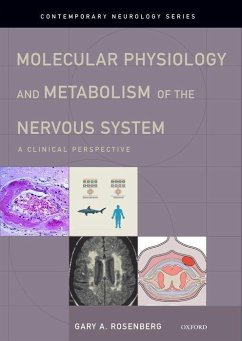
Molecular Physiology and Metabolism of the Nervous System
Gary A Rosenberg
Molecular Physiology and Metabolism of the Nervous System
- Gebundenes Buch
- Merkliste
- Auf die Merkliste
- Bewerten Bewerten
- Teilen
- Produkt teilen
- Produkterinnerung
- Produkterinnerung
This book, authored by Gary A. Rosenberg, an authority on the physiology of brain fluids and metabolism, combines the classic physiology that dates back to the beginning of the nineteenth century with the advances in molecular sciences, providing a strong framework for understanding the diseases that are commonly treated by neurologists.
Andere Kunden interessierten sich auch für
![Immediate Early Genes and Inducible Transcription Factors in Mapping of the Central Nervous System Function and Dysfunction Immediate Early Genes and Inducible Transcription Factors in Mapping of the Central Nervous System Function and Dysfunction]() L. Kaczmarek / H.A. Robertson (eds.)Immediate Early Genes and Inducible Transcription Factors in Mapping of the Central Nervous System Function and Dysfunction406,99 €
L. Kaczmarek / H.A. Robertson (eds.)Immediate Early Genes and Inducible Transcription Factors in Mapping of the Central Nervous System Function and Dysfunction406,99 €![Exploring the Nervous System Exploring the Nervous System]() Robert L SchoenfeldExploring the Nervous System48,99 €
Robert L SchoenfeldExploring the Nervous System48,99 €![Systemic Hereditary Degenerative and Dystrophic Diseases of the Nervous and Muscular System Systemic Hereditary Degenerative and Dystrophic Diseases of the Nervous and Muscular System]() Ivan ManchevSystemic Hereditary Degenerative and Dystrophic Diseases of the Nervous and Muscular System22,99 €
Ivan ManchevSystemic Hereditary Degenerative and Dystrophic Diseases of the Nervous and Muscular System22,99 €![Plasticity in the Human Nervous System Plasticity in the Human Nervous System]() Plasticity in the Human Nervous System100,99 €
Plasticity in the Human Nervous System100,99 €![Nerves and Nerve Injuries Nerves and Nerve Injuries]() Nerves and Nerve Injuries191,99 €
Nerves and Nerve Injuries191,99 €![Central Nervous System Infections, an Issue of Neuroimaging Clinics Central Nervous System Infections, an Issue of Neuroimaging Clinics]() Guarang ShahCentral Nervous System Infections, an Issue of Neuroimaging Clinics119,99 €
Guarang ShahCentral Nervous System Infections, an Issue of Neuroimaging Clinics119,99 €![Case Studies in Stroke Case Studies in Stroke]() Michael G HennericiCase Studies in Stroke136,99 €
Michael G HennericiCase Studies in Stroke136,99 €-
-
-
This book, authored by Gary A. Rosenberg, an authority on the physiology of brain fluids and metabolism, combines the classic physiology that dates back to the beginning of the nineteenth century with the advances in molecular sciences, providing a strong framework for understanding the diseases that are commonly treated by neurologists.
Hinweis: Dieser Artikel kann nur an eine deutsche Lieferadresse ausgeliefert werden.
Hinweis: Dieser Artikel kann nur an eine deutsche Lieferadresse ausgeliefert werden.
Produktdetails
- Produktdetails
- Verlag: Hurst & Co.
- Seitenzahl: 240
- Erscheinungstermin: 30. April 2012
- Englisch
- Abmessung: 257mm x 183mm x 20mm
- Gewicht: 771g
- ISBN-13: 9780195394276
- ISBN-10: 0195394275
- Artikelnr.: 35055795
- Herstellerkennzeichnung
- Libri GmbH
- Europaallee 1
- 36244 Bad Hersfeld
- gpsr@libri.de
- Verlag: Hurst & Co.
- Seitenzahl: 240
- Erscheinungstermin: 30. April 2012
- Englisch
- Abmessung: 257mm x 183mm x 20mm
- Gewicht: 771g
- ISBN-13: 9780195394276
- ISBN-10: 0195394275
- Artikelnr.: 35055795
- Herstellerkennzeichnung
- Libri GmbH
- Europaallee 1
- 36244 Bad Hersfeld
- gpsr@libri.de
Gary A. Rosenberg, MD Chairman of Neurology Professor of Neurology, Neurosciences, Cell Biology and Physiology, and Mathematics and Statistics University of New Mexico Health Sciences Center Albuquerque, NM
* Part I: Physiology of brain fluids and blood-brain barrier
* Chapter 1: Anatomy of Fluid Interfaces that Protect the
Microenvironment
* 1.1. Historical perspective
* 1.2 Cerebral microenvironment
* 1.3. Development of the brain-fluid interfaces
* 1.3.1. Neural tube, ependymal cells and stem cells
* 1.3.2. Cilated ependymal cells and CSF movement
* 1.3.3. Choroid plexuses, arachnoid and capillaries
* 1.4. Extracellular Space and Extracellular Matrix
* 1.5. Brain-Fluid Interfaces
* 1.5.1. Anatomy of the cerebral blood vessels
* 1.5.2. Brain cells interfaces with CSF at ependymal and pia
* 1.6. Dura, arachnoid and pial layers
* 1.7. What are sources of energy?
* Chapter 2: Physiology of the Cerebrospinal and Interstitial Fluids
* 2.1. Introduction
* 2.2. Proteins in the CSF
* 2.3. CSF Pressure Reflects Venous Pressure in the Right Heart
* 2.4. Formation, Circulation and Absorption of CSF
* 2.4.1. Formation of CSF by choroid plexuses
* 2.4.2. Choroid plexus and disease biomarkers in CSF
* 2.4.3. Absorption of CSF at the arachnoid villi
* 2.5. Electrolyte balance in the CSF
* 2.6. Meninges and sites of masses and infection
* 2.7. Interstitial fluid
* 2.8. Lyphatic drainage
* 2.9. Water diffusion, bulk flow if ISH and diffusion tensor imaging
* 2.10. Neuropeptides and fluid homeostasis
* 2.11. Aquaporins and water transport in the CNS
* Chapter 3: Neurovascular Unit
* 3.1. Early experiments on blood-brain barrier
* 3.2. The Neurovascular unit and tight junction proteins
* 3.3. Integrins, selectins and endothelial cell adhesion
* 3.4. Astrocytes, pericytes and basal lamina
* 3.5. Movement of substances into and out of brain
* 3.6. Glucose and amino acid transport
* 3.7. Proteases and the neurovascular unit
* 3.8. Matrix metalloproteinases (MMPs)
* 3.9. A disintegrin and metalloproteinase (ADAM)
* 3.10. Barrier systems evolved to an endothelial barrier
* Part II: Metabolism, disorders of brain fluids, and mathematics of
transport
* Chapter 4: Glucose, Amino acid and Lipid Metabolism
* 4.1. Glucose metabolism
* 4.2. Amino acid neurotransmitters
* 4.3. Lipid metabolism
* 4.4. Eicosanoid metabolism
* 4.5. Hepatic encephalopathy
* 4.6. Hypoglycemia
* 4.7. Hyponatremia, osmotic demyelination and acid balance
* 4.7.1. Hyponatremia
* 4.7.2. Hyperglycemia
* 4.7.3. Acidosis
* Chapter 5: Disorders of Cerebrospinal Circulation: Idiopathic
Intracranial Hypertension (IIH) and Hydrocephalus
* 5.1. Introduction
* 5.2. Clinical Features of IIH
* 5.3. Treatment of IIH
* 5.4. Hydrocephalus
* 5.5. Hydrocephalus in children
* 5.6. Adult-onset hydrocephalus
* 5.6.1. Obstructive hydrocephalus
* 5.6.2. Normal-pressure hydrocephalus
* Chapter 6: Quantification of Cerebral Blood Flow and Blood Brain
Barrier Transport by NMR and PET
* 6.1. Introduction
* 6.2. Mathematical approach to cerebral blood flow and transport
* 6.2.1. Cerebral blood flow: Schmidt-Kety approach
* 6.2.2. Regional blood flow
* 6.2.3. Transport between blood and brain
* 6.3 Positron emission tomography (PET)
* 6.3.1. Single-injection external registration
* 6.3.2. Patlak graphical BBB method for autoradiography and MRI
* 6.4 Magnetic resonance imaging and spectroscopy
* 6.4.1. Multinuclear NMR
* 6.4.2. Relaxation phenomenon and the rotating frame
* 6.4.3. 31P-MRS
* 6.4.4. 13C-MRS
* 6.4.5. 1H-MRS
* Part III: Ischemia, edema and inflammation
* Chapter 7: Mechanisms of Ischemic/Hypoxic Brain Injury
* 7.1. Epidemiology, risk factors and prevention of stroke
* 7.2. Molecular cascades in ischemic tissue results from energy
failure
* 7.3. Excitatory and inhibitory neurotransmitters
* 7.4. Neuroinflammation in stroke
* 7.5. Proteases in hypoxia/ischemia
* 7.6. Caspases and cell death
* 7.7. Tissue inhibitors of metalloproteinases (TIMPs) and apoptosis
* 7.8. Tight junction proteins and MMPs
* 7.9. MMPs and tPA-induced bleeding
* 7.10. Animal models in stroke
* 7.11. Arteriovenous malformations and cavernous hemangiomas
* 7.12. MRI, PET and EPR in hypoxia-ischemia
* 7.12.1. MRI and MRS
* 7.12.2. Positron emission tomography (PET)
* 7.12.3. Electron paramagnetic resonance
* Chapter 8: Vascular Cognitive Impairment and Alzheimer's Disease
* 8.1. Regulation of cerebral blood flow
* 8.2. Hypoxia-ischemia in cardiac arrest
* 8.2.1 Prognosis for recovery after cardiac arrest
* 8.2.2 Cardiac surgery and memory loss
* 8.2.3 Delayed post anoxic leukoencephalopathy
* 8.3. Hypoxia inducible factors and gene expression
* 8.4. Intermittent hypoxia is a strong stimulus for HIF
* 8.5. Vascular cognitive impairment
* 8.6. White matter hyperintensities on MRI and Binswanger's disease
* 8.7. Alzheimer's disease, vascular disease and the amyloid hypothesis
* Chapter 9: Effects of Altitude on the Brain
* 9.1. Introduction
* 9.2. Genetic tolerance to altitude
* 9.3. Acute mountain sickness and high altitude pulmonary edema
* 9.4. High altitude cerebral edema
* 9.5. Cognitive consequences of hypobaric hypoxia
* 9.6. Imaging of the brain at high altitude
* 9.7. Hypoxia-inducible factors and sleep disorders in AMS
* 9.8. Treatment of altitude illnesses
* Chapter 10: Brain Edema
* 10.1. Introduction
* 10.2. Role of aquaporins in brain edema
* 10.3. Role of Neuroinflammation in the formation of vasogenic edema
* 10.3.1. Oxidative stress and brain edema
* 10.3.2 . Arachidonic acid and brain edema
* 10.3.3. Vascular endothelial growth factor and angiopoietins
* 10.4. Clinical conditions associated with brain edema
* 10.5. Imaging brain edema
* 10.6 . Treatment of brain edema and hypoxic/ischemic injury
* 10.7. Multiple drugs for treatment of ischemia
* Chapter 11: Intracerebral Hemorrhage
* 11.1. Introduction
* 11.2. History of ICH
* 11.3. Molecular mechanisms in ICH
* 11.4. Clinical aspects of intracranial bleeding
* 11.5. Pathophysiology of ICH: Evidence from animal studies
* 11.6 Extrapolation of experimental results to treatments for ICH
* Chapter 12: Autoimmunity, Hypoxia, and Inflammation in Demyelinating
Diseases
* 12.1. Introduction
* 12.2. Heterogeneity of the pathological findings in MS
* 12.3. Proteases implicated in MS pathology
* 12.4. BBB disruption in MS
* 12.5. Devic's neuromyelitis optica
* 12.6. Nonimmunological processes in demyelination
* 12.7. Experimental allergic encephalomyelitis and pathogenesis of MS
* 12.8. Epilogue- synthesis and future directions

