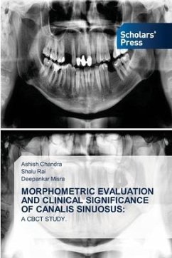
The Canalis Sinuosus (CS) is a neurovascular canal, through which the Anterior Superior Alveolar Nerve (ASAN) passes and then curves medially in path between the nasal cavity and the maxillary sinus, reaching the premaxilla in the canine to incisor region. The purpose of this article is to report two cases with the presence of Canalis Sinuosus, in order to alert and guide professionals and discuss the morphology of this anatomical variation avoiding postsurgical disorders. The cases revealed the presence of Canalis Sinuosus in Cone Beam Computed Tomography (CBCT) imaging. The knowledge of this anatomical variation is essential for professionals, because attention to this region prevents irreversible damage. Therefore, the use of advanced imaging is recommended during the treatment planning stages and in patients undergoing surgery treatment and after surgery in this area.Keyword: Canalis Sinuosus (CS), Anterior Superior Alveolar Nerve (ASAN), Cone Beam Computed Tomography (CBCT),Advanced Imaging, Anterior Maxilla, Maxillary Surgery.
Bitte wählen Sie Ihr Anliegen aus.
Rechnungen
Retourenschein anfordern
Bestellstatus
Storno

