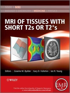
The content of this volume has been added to eMagRes (formerly Encyclopedia of Magnetic Resonance) - the ultimate online resource for NMR and MRI. Up to now MRI could not be used clinically for imaging fine structures of bones or muscles. Since the late 1990s however, the scene has changed dramatically. In particular, Graeme Bydder and his many collaborators have demonstrated the possibility - and importance - of imaging structures in the body that were previously regarded as being "MR Invisible". The images obtained with a variety of these newly developed methods exhibit complex contrast, resulting in a new quality of images for a wide range of new applications. This Handbook is designed to enable the radiology community to begin their assessment of how best to exploit these new capabilities. It is organised in four major sections - the first of which, after an Introduction, deals with the basic science underlying the rest of the contents of the Handbook. The second, larger, section describes the techniques which are used in recovering the short T2 and T2* data from which the images are reconstructed. The third and fourth sections present a range of applications of the methods described earlier. The third section deals with pre-clinical uses and studies, while the final section describes a range of clinical applications. It is this last section that will surely have the biggest impact on the development in the next few years as the huge promise of Short T2 and T2* Imaging will be exploited to the benefit of patients. In many instances, the authors of an article are the only research group who have published on the topic they describe. This demonstrates that this Handbook presents a range of methods and applications with a huge potential for future developments. About EMR Handbooks / eMagRes Handbooks The Encyclopedia of Magnetic Resonance (up to 2012) and eMagRes (from 2013 onward) publish a wide range of online articles on all aspects of magnetic resonance in physics, chemistry, biology and medicine. The existence of this large number of articles, written by experts in various fields, is enabling the publication of a series of EMR Handbooks / eMagRes Handbooks on specific areas of NMR and MRI. The chapters of each of these handbooks will comprise a carefully chosen selection of articles from eMagRes. In consultation with the eMagRes Editorial Board, the EMR Handbooks / eMagRes Handbooks are coherently planned in advance by specially-selected Editors, and new articles are written (together with updates of some already existing articles) to give appropriate complete coverage. The handbooks are intended to be of value and interest to research students, postdoctoral fellows and other researchers learning about the scientific area in question and undertaking relevant experiments, whether in academia or industry. Have the content of this Handbook and the complete content of eMagRes at your fingertips! Visit: www.wileyonlinelibrary.com/ref/eMagRes View other eMagRes publications here
Bitte wählen Sie Ihr Anliegen aus.
Rechnungen
Retourenschein anfordern
Bestellstatus
Storno

