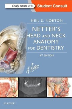
A concise and visual guide to clinically relevant anatomy for dentistry, Netter's Head and Neck Anatomy for Dentistry is an effective text for class and exam preparation, as well as a quick review in professional practice. Concise text, high-yield tables, clinical correlations, and review questions combine to make this new edition a perfect choice for learning and remembering the need-to-know structures, relationships, and concepts, while beautiful illustrations created in the Netter tradition enhance your visual mastery of the material.
You may also be interested in:
A companion set of flash cards, Netter's Advanced Head & Neck Anatomy Flash Cards, 3rd Edition.
Over 100 multiple-choice questions complete with explanations help you assess your knowledge of the material and prepare for exams. Identify clinically relevant anatomy with Netter illustrations and new art created in the Netter tradition. Concise text and high-yield tables offer fast access to important facts. Procedures coverage gives context and clinical meaning to the anatomy. Expanded, up-to-date coverage on dental implants, cone beam imaging, and mandible osteology. Beautiful new illustrations by Carlos Machado, MD, of the TMJ, articular disc pathology, infratemporal fossa, pterygopalatine fossa, and maxillary artery. Interactive eBook included with print purchase, which includes access to the full text, interactive images, case studies, additional assessment questions, video clips from cone beam CTs, and a rotatable 3D skull.
Hinweis: Dieser Artikel kann nur an eine deutsche Lieferadresse ausgeliefert werden.


