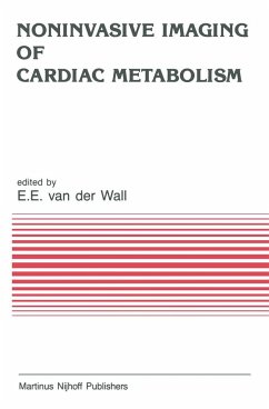
F.J.Th. WACKERS Metabolic imaging: The future of cardiovascular nuclear imaging? Since cardiovascular nuclear imaging emerged as a new subspecialty in the mid-1970s, the field has gone through an explosive growth. Radionuclide techniques became readily recognized as important new diagnostic aids in the armamentarium of the clinical cardiologist. Initially, cardiovascular nuclear imaging focused on static myocardial imaging using either thallium-201 or technetium-99m-pyrophosphate for diagnosing acute myocardial infarction. Shortly thereafter, multigated equilibrium radionuclide angiocardiography became the most widely used noninvasive method for assessing cardiac function. Furthermore, attention and clinical application shifted towards the use of radionuclide techniques in conjunction with exercise testing, either with thallium-20 1 myocardial perfusion imaging or technetium-99m left ventricular function studies. The future of cardiovascular nuclear imaging appeared exciting and promising. However, around 1980 pessimists predicted the premature demise of cardiovascular nuclear imaging with the introduction of digital subtraction angiography and nuclear magnetic resonance imaging. These doomsayers have been proven wrong: in 1985 cardiovascular nuclear imaging is thriving and, in many centers, even expanding. Although digital substraction angiography and magnetic resonance imaging provided exquisite anatomic detail, for practical evaluation of patients with ischemic heart disease - in the Coronary Care Unit or exercise laboratory - nuclear techniques appeared to be more practical.
Hinweis: Dieser Artikel kann nur an eine deutsche Lieferadresse ausgeliefert werden.
Hinweis: Dieser Artikel kann nur an eine deutsche Lieferadresse ausgeliefert werden.

