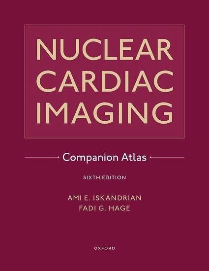
Nuclear Cardiac Imaging Companion Atlas
Herausgeber: Iskandrian, Ami E; Hage, Fadi G
Nuclear Cardiac Imaging Companion Atlas
Herausgeber: Iskandrian, Ami E; Hage, Fadi G
- Gebundenes Buch
- Merkliste
- Auf die Merkliste
- Bewerten Bewerten
- Teilen
- Produkt teilen
- Produkterinnerung
- Produkterinnerung
A companion to the 6th edition Nuclear Cardiac Imaging, the atlas provides an expansive set of case examples and information not included in the textbook. An abundance of multiple-choice exam questions throughtout serves as a challenging self-assessment tool for readers testing their knowledge.
Andere Kunden interessierten sich auch für
- Arvind N. ShuklaBIOMEDICAL IMAGING61,99 €
- Narang Karanjit SinghINTRAOPERATIVE IMAGING IN NEUROSURGERY94,99 €
- Robert J. SeidelFrom Principles of Learning to Strategies for Instruction-With Workbook Companion62,99 €
- Titus StrongThe Young Scholar's Manual, Or Companion to the Spelling Book Consisting of Easy Lessons in the Several Branches of Early Education33,99 €
- The Routledge Companion to Dyslexia317,99 €
- Robin AlexanderA Dialogic Teaching Companion227,99 €
- Parimala V. RaoThe Routledge Companion to the History of Education in India, 1780-1947235,99 €
-
-
-
A companion to the 6th edition Nuclear Cardiac Imaging, the atlas provides an expansive set of case examples and information not included in the textbook. An abundance of multiple-choice exam questions throughtout serves as a challenging self-assessment tool for readers testing their knowledge.
Hinweis: Dieser Artikel kann nur an eine deutsche Lieferadresse ausgeliefert werden.
Produktdetails
- Produktdetails
- Verlag: Oxford University Press
- Seitenzahl: 240
- Erscheinungstermin: 16. Februar 2024
- Englisch
- Abmessung: 239mm x 226mm x 23mm
- Gewicht: 1021g
- ISBN-13: 9780197521434
- ISBN-10: 0197521436
- Artikelnr.: 67734319
- Herstellerkennzeichnung
- Libri GmbH
- Europaallee 1
- 36244 Bad Hersfeld
- gpsr@libri.de
Ami E. Iskandrian is a Distinguished Professor of Medicine and Radiology in the Division of Cardiovascular Disease at the University of Alabama at Birmingham. University's School of Medicine in Atlanta, Georgia. Fadi G. Hage is a Professor of Medicine in the Division of Cardiovascular Disease at the University of Alabama at Birmingham. He is the Chief of Cardiology at the Birmingham VA Medical Center and the Director of Nuclear Cardiology at the University of Alabama at Birmingham. Fadi G. Hage is a Professor of Medicine in the Division of Cardiovascular Disease at the University of Alabama at Birmingham. He is the Chief of Cardiology at the Birmingham VA Medical Center and the Director of Nuclear Cardiology at the University of Alabama at Birmingham.
* Section I. Historical, Technical and Physiological Considerations
* 1. Nuclear Cardiology: History and Milestones
* 2. Radiation physics
* 3. SPECT and PET Instrumentation
* 4. Kinetics of Conventional and New Cardiac Radiotracers
* 5. Radionuclide Angiography: Planar and Tomographic
* 6. Gated SPECT MPI: Imaging Protocols and Acquisition
* 7. Image Artifacts
* Section II. Diagnosis and Risk Assessment
* 8. Regulation of Myocardial Blood Flow
* 9. How to measure Myocardial Blood Flow by PET?
* 10. How to measure Myocardial Blood flow by SPECT?
* 11. Treadmill Exercise Testing
* 12. Pharmacological Stress Testing
* 13. SPECT MPI in Diagnosis/Risk Assessment
* 14. SPECT MPI for Risk Assessment in special groups (DM, Kidney
transplant, Liver transplant, Asymptomatic, Obese)
* 15. SPECT MPI for Risk Assessment before non-cardiac surgery
* 16. Evaluation of patients with Chest Pain in the Emergency
Department
* 17. Role of PET (Perfusion and MBF) in Diagnosis and Risk Assessment
* 18. Nuclear Imaging in women
* 19. Myocardial Viability Assessment by Nuclear Techniques
* Section III. Role of Nuclear Imaging Beyond CAD
* 20. Imaging dyssynchrony
* 21. Imaging Myocardial Innervation by SPECT and PET
* 22. Imaging sarcoid heart disease
* 23. Imaging Amyloid heart disease
* 24. Imaging infections of valves and devices
* 25. Imaging cardiac and vascular inflammation
* 26. Imaging in Heart Failure including those with devices and post
cardiac transplant
* Section IV. Advances in Nuclear Cardiac Imaging
* 27. Evolving role of CT, cMR and echo in CAD
* 28. Hybrid Imaging: SPECT/CTA, PET/MR and SPECT Calcium Score: when
and why?
* 29. Artificial Intelligence and nuclear imaging
* 30. Nuclear Imaging in patients with serious arrhythmias
* 31. Nuclear Imaging in patients with HIV
* 32. Nuclear Imaging in cardio-oncology
* 33. Nuclear Imaging in patients with congenital heart diseases
* Section V. Challenges for Nuclear Cardiology
* 34. Physician Certification and Lab Accreditation
* 35. ACC/AHA/ESC Nuclear Guidelines and Appropriate Use Criteria
* 36. Nuclear Cardiology Report generation
* 37. Radiation Considerations in imaging
* 38. Statistics in Nuclear imaging
* 39. Nuclear Imaging in Developing Countries
* 40. Ask the Experts
* Section VI. Self-Assessment
* 41. Nuclear Cardiology Self-Assessment