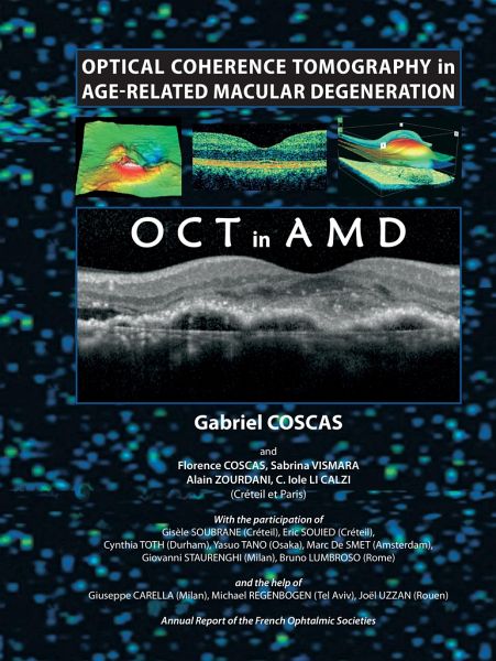
Optical Coherence Tomography in Age-Related Macular Degeneration

PAYBACK Punkte
76 °P sammeln!
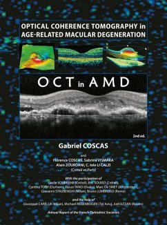
Rapid or even dramatic progress has been made in the field of AMD over recent years, leading to a constant revision of basic concepts. A wide range of fundus imaging modalities are now available, and this book explains the respective value of each technique. The information provided by OCT is presented logically by comparison with plain films, autofluorescence, fluorescein angiography, or indocyanine green angiography. Meticulous biomicroscopic examination of macular changes and the essential value of fluorescein angiography for the detection of anatomical alterations of the macula and for pre...
Rapid or even dramatic progress has been made in the field of AMD over recent years, leading to a constant revision of basic concepts. A wide range of fundus imaging modalities are now available, and this book explains the respective value of each technique. The information provided by OCT is presented logically by comparison with plain films, autofluorescence, fluorescein angiography, or indocyanine green angiography. Meticulous biomicroscopic examination of macular changes and the essential value of fluorescein angiography for the detection of anatomical alterations of the macula and for precise evaluation of lesions and their course by indocyanine green angiography have naturally led the author Gabriel Coscas to analyze the new data provided by OCT.


