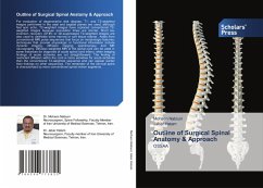
For evaluation of degenerative disk disease, T1- and T2-weighted images performed in the axial and sagittal planes are used, although fast spin echo, T2-weighted images have replaced conventional T2-weighted images because acquisition times are shorter. Short tau inversion recovery (STIR) or fat-suppressed T2-weighted images are also used to delineate bone marrow or soft tissue edema . Aside from conventional MRI pulse sequences that focus on morphologic features, techniques that provide physiologic or functional information include dynamic imaging, diffusion imaging, spectroscopy, and MR neurography. Diffusion-weighted MRI of the spinal cord can be used in some instances for diagnosis of acute spinal cord infarct, when imaging findings of acute ischemia are not straightforward. The finding of restricted diffusion within the cord is more sensitive for acute ischemia than the conventional T2-weighted sequence and can appear earlier than findings on other sequences. The remainder of the cervical spine is characterized by more conventional spinal motion segments .
Bitte wählen Sie Ihr Anliegen aus.
Rechnungen
Retourenschein anfordern
Bestellstatus
Storno

