Photography in Orthodontics
Versandkostenfrei!
Versandfertig in 6-10 Tagen
40,99 €
inkl. MwSt.

PAYBACK Punkte
20 °P sammeln!
Photographs of a patient's face play a crucial role in guiding diagnosis and treatment planning in dentistry, providing valuable documentation for preoperative and post-treatment conditions. Unlike cephalometry, which primarily captures soft tissues in profile, photographs offer a more conventional and visually accessible method for monitoring dentofacial changes during growth. They serve as reliable qualitative tools for evaluating postoperative results, enabling patients to review their transformations. In the realm of orthodontics and other dental disciplines, the demand for photo-documenta...
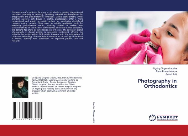
Photographs of a patient's face play a crucial role in guiding diagnosis and treatment planning in dentistry, providing valuable documentation for preoperative and post-treatment conditions. Unlike cephalometry, which primarily captures soft tissues in profile, photographs offer a more conventional and visually accessible method for monitoring dentofacial changes during growth. They serve as reliable qualitative tools for evaluating postoperative results, enabling patients to review their transformations. In the realm of orthodontics and other dental disciplines, the demand for photo-documentation is on the rise. The advent of digital photography in clinical settings is generating excitement, offering the potential for cost-effective, high-quality imaging with the integration of computer technology. The continuous evolution of technology in dentistry is evident, opening new possibilities for improved patient care and research.


