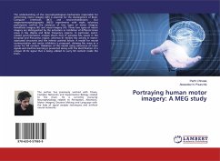
Portraying human motor imagery: A MEG study
Versandkostenfrei!
Versandfertig in 6-10 Tagen
27,99 €
inkl. MwSt.

PAYBACK Punkte
14 °P sammeln!
The understanding of the neurophysiological mechanisms responsible for performing motor imagery (MI) is essential for the development of Brain-Computer Interfaces (BCI) and neurorehabilitation. Our magnetoencephalographic (MEG) experiments with eight voluntary participants confirm the existence of two types of motor imagery: kinesthetic imagery (KI) and visual imagery (VI). These two types of motor imagery are distinguished by the activation or inhibition of different brain areas in Mu (Alpha and Beta) frequency regions. In particular, event-related synchronization analysis shows that VI activ...
The understanding of the neurophysiological mechanisms responsible for performing motor imagery (MI) is essential for the development of Brain-Computer Interfaces (BCI) and neurorehabilitation. Our magnetoencephalographic (MEG) experiments with eight voluntary participants confirm the existence of two types of motor imagery: kinesthetic imagery (KI) and visual imagery (VI). These two types of motor imagery are distinguished by the activation or inhibition of different brain areas in Mu (Alpha and Beta) frequency regions. In particular, event-related synchronization analysis shows that VI activates Mu waves in the Occipital and Precuneus region, whereas KI inhibits Mu activity in motor-associated structures and the inferior parietal lobule. A model for neural communication and motor inhibition is proposed, claiming Mu wave as a carrier for MI content. Validation of this model using coherence of brain signals and machine learning is presented along with the identification of a unique 45 Hz signal that is being utilized to carry MI content inside the brain.


