• Produktbild: Practical Scanning Electron Microscopy
  • Produktbild: Practical Scanning Electron Microscopy

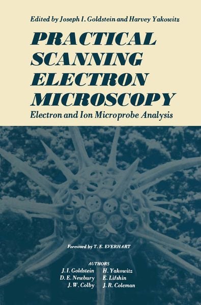
Practical Scanning Electron Microscopy Electron and Ion Microprobe Analysis

97,99 €

inkl. gesetzl. MwSt., Versandkostenfrei


Beschreibung

Produktdetails

Einband

Taschenbuch

Erscheinungsdatum

12.10.2011

Abbildungen

XVIII, 582 p.

Herausgeber

Joseph Goldstein

Verlag

Springer Us

Seitenzahl

582

Maße (L/B/H)

23,5/15,5/3,3 cm

Gewicht

902 g

Auflage

Softcover reprint of the original 1st ed. 1975

Sprache

Englisch

ISBN

978-1-4613-4424-7

Beschreibung

Portrait

Joseph Goldstein leitet seit 1974 weltweit Retreats und Seminare in buddhistischer Meditation und ist Mitbegründer der Insight Meditation Society.
Seine Praxis begann er 1967, als er als Friedensaktivist in Thailand auf den Buddhismus stieß. Heute gilt er als einer der führenden Vertreter eines westlichen Buddhismus. In deutscher Sprache wurde von ihm (gemeinsam mit Jack Kornfield) das Buch Einsicht durch Meditation veröffentlicht.

Produktdetails

Einband

Taschenbuch

Erscheinungsdatum

12.10.2011

Abbildungen

XVIII, 582 p.

Herausgeber

Joseph Goldstein

Verlag

Springer Us

Seitenzahl

582

Maße (L/B/H)

23,5/15,5/3,3 cm

Gewicht

902 g

Auflage

Softcover reprint of the original 1st ed. 1975

Sprache

Englisch

ISBN

978-1-4613-4424-7

Herstelleradresse

Springer-Verlag KG
Sachsenplatz 4-6
1201 Wien
AT

Email: ProductSafety@springernature.com

Noch keine Bewertungen vorhanden

Verfassen Sie die erste Bewertung zu diesem Artikel

Helfen Sie anderen Kundinnen und Kunden durch Ihre Meinung.

Kundinnen und Kunden meinen

Bewertungen (0)

  • Produktbild: Practical Scanning Electron Microscopy
  • Produktbild: Practical Scanning Electron Microscopy
  • I Introduction.- I. Evolution of the Scanning Electron Microscope.- II. Evolution of the Electron Probe Microanalyzer.- III. Combination SEM-EPMA.- IV. Outline and Purpose of This Book.- References.- Bibliography of Texts and Monographs in SEM and EPMA.- II Electron Optics.- I. Electron Guns.- II. Electron Lenses.- III. Electron Probe Diameter dp vs. Electron Probe Current i.- IV. Depth of Field.- References.- III Electron Beam-Specimen Interaction.- I. Electron Scattering in Solids.- II. Electron Range and Spatial Distribution of the Primary Electron Beam.- III. Emitted Electrons—Backscattered Electrons.- IV. Emitted Electrons—Low-Energy Electrons.- V. X-Rays.- VI. Auger Electrons.- VII. Summary—Range and Spatial Resolution.- References.- IV Image Formation in the Scanning Electron Microscope.- I. The SEM Imaging Process.- II. Signal Detectors.- III. Contrast Formation.- IV. Signal Characteristics and Image Quality.- V. Resolution Limitations in the SEM.- VI. Signal Processing.- VII. Image Defects.- VIII. Electron Penetration Effects in Images.- References.- V Contrast Mechanisms of Special Interest In Materials Science.- I. Introduction.- II. Electron Channeling Contrast.- III. Magnetic Contrast in the SEM.- IV. Voltage Contrast.- V. Electron-Beam-Induced Current (EBIC).- VI. Cathodoluminescence.- References.- VI Specimen Preparation, Special Techniques, and Applications of the Scanning Electron Microscope.- I. Specimen Preparation for Materials Examination in the SEM.- II. Stereomicroscopy.- III. Dynamic Experiments in the SEM.- IV. Applications of the SEM.- References.- VII X-Ray Spectral Measurement and Interpretation.- I. Introduction.- II. Crystal Spectrometers.- III. Solid State X-Ray Detectors.- IV. A Comparison of Crystal Spectrometers with Solid StateX-Ray Detectors.- V. The Analysis of X-Ray Spectral Data.- References.- VIII Microanalysis of Thin Films and Fine Structure.- I. Introduction.- II. Factors Affecting X-Ray Spatial Resolution.- III. Characterizing the X-Ray-Excited Volume.- IV. Thin-Film Analysis.- V. Particles, Inclusions, and Fine Structures.- References.- IX Methods of Quantitative X-Ray Analysis Used in Electron Probe Microanalysis and Scanning Electron Microscopy.- I. Introduction.- II. The Absorption Factor kA.- III. Atomic Number Correction kZ.- IV. The Characteristic Fluorescence Correction kF.- V. The Continuum Fluorescence Correction.- VI. Summary Discussion of the ZAF Method.- VII. The Empirical Method for Quantitative Analysis.- VIII. Comments on Analysis Involving Elements of Atomic Number of 11 or Less.- IX. Quantitative Analysis with Nonnormal Electron-Beam Incidence.- X. Analysis Involving Special Specimen Geometries.- XI. Discussion.- Appendix. The Analysis of an Iron-Silicon Alloy.- References.- X Computational Schemes for Quantitative X-Ray Analysis: On-Line Analysis with Small Computers.- I. Introduction.- II. Summary of Computational Schemes for Quantitative Analysis.- III. The FRAME Program.- IV. Data Reduction Based on the Hyperbolic Method.- V. Summary.- References.- XI Practical Aspects of X-Ray Microanalysis.- I. Grappling with the Unknown.- II. Specimen Preparation for Quantitative Analysis.- III. Applications Involving Compositional Analysis.- References.- XII Special Techniques in the X-Ray Analysis of Samples.- I. Light Element Analysis.- II. Precision and Sensitivity in X-Ray Analysis.- III. X-Ray Analysis at Interfaces.- IV. Soft X-Ray Emission Spectra.- V. Thin Films.- Appendix. Deconvolution Technique.- References.- XIII Biological Applications: Sample Preparation and Quantitation.- I. Sample Preparation.- II. Analysis.- III. Summary.- References.- XIV Ion Microprobe Mass Analysis.- I. Basic Concepts and Instrumentation.- II. Ion Microscope.- III. Ion Microprobe.- IV. Production of Ions.- V. Sputtering.- VI. Qualitative Analysis.- VII. Quantitative Analysis.- VIII. Dead-Time Losses.- IX. In-Depth Profiling.- X. Applications.- References.