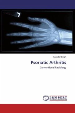
Psoriatic arthritis shows wide range of radiological skeletal changes. The most commonly sites involved are "bare area" erosions of distal interphalangeal joints of fingers and inter phalangeal joints of great toe. The most common radiological findings are erosion in hands and new bone formation in feet. In spine dorsolumbar region is commonly involved in form of coarse, asymmetrical syndemophytes. Articular erosions is a common findings in sacroiliac joints. In routine, radiological assessment is not done in psoriatic cases with or without arthritis. It is of the opinion that every patient of psoriasis must be subjected to radiological examination so as to detect arthritis at an early stage,to start early aggressive therapy and to stop the progression of joint destruction and disability.
Bitte wählen Sie Ihr Anliegen aus.
Rechnungen
Retourenschein anfordern
Bestellstatus
Storno

