Rapid Review of MRI
Versandkostenfrei!
Versandfertig in 6-10 Tagen
43,99 €
inkl. MwSt.

PAYBACK Punkte
22 °P sammeln!
Magnetic resonance imaging (MRI) allows doctors to see the inside of the human body in amazing detail using magnets and radio waves. The first MRI scanner used to image the human body was built in 1977 in New York. Since then, the technology has come a long way, and MRI is now used frequently to examine the inside of the human body. The advantage of MRI devices, unlike X-ray imaging devices or CT scans, is that radiation is not given to the body, and that is why the use of this diagnostic method has been more and more popular with specialists. The MRI scanner is a large tunnel containing power...
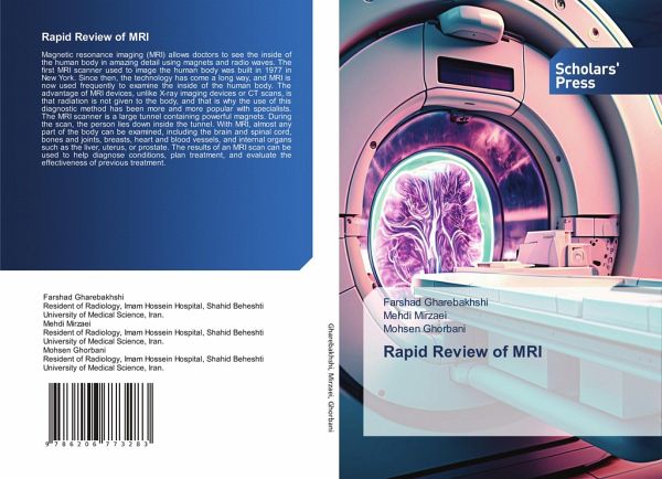
Magnetic resonance imaging (MRI) allows doctors to see the inside of the human body in amazing detail using magnets and radio waves. The first MRI scanner used to image the human body was built in 1977 in New York. Since then, the technology has come a long way, and MRI is now used frequently to examine the inside of the human body. The advantage of MRI devices, unlike X-ray imaging devices or CT scans, is that radiation is not given to the body, and that is why the use of this diagnostic method has been more and more popular with specialists. The MRI scanner is a large tunnel containing powerful magnets. During the scan, the person lies down inside the tunnel. With MRI, almost any part of the body can be examined, including the brain and spinal cord, bones and joints, breasts, heart and blood vessels, and internal organs such as the liver, uterus, or prostate. The results of an MRI scan can be used to help diagnose conditions, plan treatment, and evaluate the effectiveness of previous treatment.


