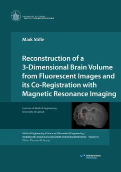
In the research of neuronal diseases, a comparison of magnetic resonance imaging (MRI) and histology sections is indispensable. While histology provides information about microscopic structures and chemical composition of brain tissue, in-vivo high-contrast MRI is a powerful tool for detecting pathologic structures in living animals. In clinic practice, the histology-MRI correspondence is often determined visually by experienced neuroscientists. This is a time-consuming and laborious procedure. In this Book an imaging pipeline in described that automatically aligns histology sections to the anatomically corresponding position in the MRI. In order to evaluate the underlying methodology, corresponding anatomical landmarks in MRI and histology were selected by experts. The accuracy achieved by the reconstruction and registration pipeline enables a precise analysis of microstructural features seen in the histology sections and superimposed on the MRI. This in turn is extremely valuable for studying the cellular mechanisms that are responsible for signal changes in MRI.
Bitte wählen Sie Ihr Anliegen aus.
Rechnungen
Retourenschein anfordern
Bestellstatus
Storno

