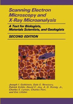
Scanning Electron Microscopy and X-Ray Microanalysis
A Text for Biologists, Materials Scientists, and Geologists
Versandkostenfrei!
Versandfertig in 1-2 Wochen
78,99 €
inkl. MwSt.
Weitere Ausgaben:

PAYBACK Punkte
39 °P sammeln!
This book has evolved by processes of selection and expansion from its predecessor, Practical Scanning Electron Microscopy (PSEM), published by Plenum Press in 1975. The interaction of the authors with students at the Short Course on Scanning Electron Microscopy and X-Ray Microanalysis held annually at Lehigh University has helped greatly in developing this textbook. The material has been chosen to provide a student with a general introduction to the techniques of scanning electron microscopy and x-ray microanalysis suitable for application in such fields as biology, geology, solid state physi...
This book has evolved by processes of selection and expansion from its predecessor, Practical Scanning Electron Microscopy (PSEM), published by Plenum Press in 1975. The interaction of the authors with students at the Short Course on Scanning Electron Microscopy and X-Ray Microanalysis held annually at Lehigh University has helped greatly in developing this textbook. The material has been chosen to provide a student with a general introduction to the techniques of scanning electron microscopy and x-ray microanalysis suitable for application in such fields as biology, geology, solid state physics, and materials science. Following the format of PSEM, this book gives the student a basic knowledge of (1) the user-controlled functions of the electron optics of the scanning electron microscope and electron microprobe, (2) the characteristics of electron-beam-sample inter actions, (3) image formation and interpretation, (4) x-ray spectrometry, and (5) quantitative x-ray microanalysis. Each of these topics has been updated and in most cases expanded over the material presented in PSEM in order to give the reader sufficient coverage to understand these topics and apply the information in the laboratory. Throughout the text, we have attempted to emphasize practical aspects of the techniques, describing those instru ment parameters which the microscopist can and must manipulate to obtain optimum information from the specimen. Certain areas in particular have been expanded in response to their increasing importance in the SEM field. Thus energy-dispersive x-ray spectrometry, which has undergone a tremendous surge in growth, is treated in substantial detail.


