Marilyn Arsham, Helen Lawce, Margaret Barch
The Agt Cytogenetics Laboratory Manual
Herausgeber: Arsham, Marilyn S; Lawce, Helen J; Barch, Margaret J
Marilyn Arsham, Helen Lawce, Margaret Barch
The Agt Cytogenetics Laboratory Manual
Herausgeber: Arsham, Marilyn S; Lawce, Helen J; Barch, Margaret J
- Gebundenes Buch
- Merkliste
- Auf die Merkliste
- Bewerten Bewerten
- Teilen
- Produkt teilen
- Produkterinnerung
- Produkterinnerung
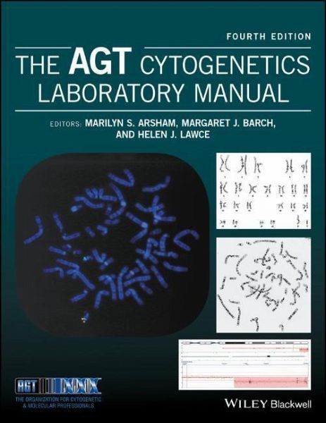
Die Zyto- oder Zellgenetik untersucht die Morphologie, Struktur, Pathologie, Funktion und das Verhalten von Chromosomen. Entstanden ist dieses Teilgebiet der Genetik, um zytogenetische Veränderungen auf Molekularebene abzubilden. Heute spricht man in diesem Zusammenhang von der Zellgenomik. Zellgenetiker greifen auf eine ganze Reihe von Verfahren zurück, um Chromosomen und/oder eine Zielregion eines bestimmten Chromosom in der Meta- oder Interphase zu untersuchen. Zu den Tools gehören Routineanalysen von Chromosomen (G-Banding), spezielle Stains für bestimmte Chromosomenstrukturen und…mehr
Andere Kunden interessierten sich auch für
- Natural Genetic Engineering and Natural Genome Editing, Volume 1178166,99 €
- Pasternak, Jack, K.An Introduction to Human Molecular Genetics185,99 €
- E O WileyPhylogenetics126,99 €
- Cassidy and Allanson's Management of Genetic Syndromes275,99 €
- Thomas LiehrBenign and Pathological Chromosomal Imbalances107,99 €
- Jeremy W. DaleFrom Genes to Genomes57,99 €
- Toren Finkel / J. Silvio Gutkind (Hgg.)Signal Transduction and Human Disease262,99 €
-
-
-
Die Zyto- oder Zellgenetik untersucht die Morphologie, Struktur, Pathologie, Funktion und das Verhalten von Chromosomen. Entstanden ist dieses Teilgebiet der Genetik, um zytogenetische Veränderungen auf Molekularebene abzubilden. Heute spricht man in diesem Zusammenhang von der Zellgenomik.
Zellgenetiker greifen auf eine ganze Reihe von Verfahren zurück, um Chromosomen und/oder eine Zielregion eines bestimmten Chromosom in der Meta- oder Interphase zu untersuchen. Zu den Tools gehören Routineanalysen von Chromosomen (G-Banding), spezielle Stains für bestimmte Chromosomenstrukturen und molekulare Sonden, z. B. im Bereich der Fluoreszenz-in-situ-Hybridisierung (FISH) sowie der Chromosomenanalyse auf Microarray-Basis. Zum Einsatz kommen eine Vielzahl von Methoden, um eine zu untersuchenden Region hervorzuheben, der so klein ist, wie eine einzige, spezifische Gensequenz.
Hinweis: Dieser Artikel kann nur an eine deutsche Lieferadresse ausgeliefert werden.
Zellgenetiker greifen auf eine ganze Reihe von Verfahren zurück, um Chromosomen und/oder eine Zielregion eines bestimmten Chromosom in der Meta- oder Interphase zu untersuchen. Zu den Tools gehören Routineanalysen von Chromosomen (G-Banding), spezielle Stains für bestimmte Chromosomenstrukturen und molekulare Sonden, z. B. im Bereich der Fluoreszenz-in-situ-Hybridisierung (FISH) sowie der Chromosomenanalyse auf Microarray-Basis. Zum Einsatz kommen eine Vielzahl von Methoden, um eine zu untersuchenden Region hervorzuheben, der so klein ist, wie eine einzige, spezifische Gensequenz.
Hinweis: Dieser Artikel kann nur an eine deutsche Lieferadresse ausgeliefert werden.
Produktdetails
- Produktdetails
- Verlag: Wiley & Sons
- Artikelnr. des Verlages: 1W119061220
- 4. Aufl.
- Seitenzahl: 1168
- Erscheinungstermin: 24. April 2017
- Englisch
- Abmessung: 287mm x 220mm x 53mm
- Gewicht: 3544g
- ISBN-13: 9781119061229
- ISBN-10: 1119061229
- Artikelnr.: 43918439
- Verlag: Wiley & Sons
- Artikelnr. des Verlages: 1W119061220
- 4. Aufl.
- Seitenzahl: 1168
- Erscheinungstermin: 24. April 2017
- Englisch
- Abmessung: 287mm x 220mm x 53mm
- Gewicht: 3544g
- ISBN-13: 9781119061229
- ISBN-10: 1119061229
- Artikelnr.: 43918439
About the Editors Marilyn S. Arsham, (retired) Cytogenetic Technologist II, Western Connecticut Health Network, Danbury Hospital campus, Danbury, Connecticut, USA. Margaret J. Barch, (formerly) Frank F Yen Cytogenetics Laboratory, Weisskopf Child Evaluation Center, University of Louisville, USA. Helen J. Lawce, Clinical Cytogenetics, Oregon Health & Science University Knight Diagnostics Laboratory, USA.
Contributing authors xxvii
Preface xxix
Acknowledgments xxxi
1 The cell and cell division 1
Margaret J. Barch and Helen J. Lawce
1.1 The cell 1
1.2 The cell cycle 14
1.3 Recombinant DNA techniques 19
1.4 The human genome 21
References 22
2 Cytogenetics: an overview 25
Helen J. Lawce and Michael G. Brown
2.1 Introduction 25
2.2 History of human cytogenetics 25
2.3 Cytogenetics methods 29
2.4 Slide-making 49
2.5 Chromosome staining 58
2.6 Chromosome microscopy/analysis 59
2.7 Laboratory procedure manual 69
References 70
Contributed protocols 75
Protocol 2.1 Slide-making 75
Protocol 2.2 Slide-making 76
Protocol 2.3 Making wet slides for chromosome analysis 78
Protocol 2.4 Slide-making 82
Protocol 2.5 Slide preparation 82
Protocol 2.6 Slide preparation procedure 84
3 Peripheral blood cytogenetic methods 87
Helen J. Lawce and Michael G. Brown
3.1 Using peripheral blood for cytogenetic analysis 87
3.2 Special uses of peripheral blood cultures 88
3.3 Peripheral blood constituents 89
3.4 Specimen handling 91
3.5 Cell culture equipment and supplies 93
3.6 Harvesting peripheral blood cultures 95
3.7 Chromosome analysis of peripheral blood 95
3.8 Storage of fixed specimens 95
Acknowledgments 95
References 95
Contributed protocols 98
Protocol 3.1 Blood culture and harvest procedure 98
Protocol 3.2 High-resolution peripheral blood method 100
Protocol 3.3 Constitutional cytogenetic studies on peripheral blood 108
Protocol 3.4 Blood culture and harvest procedure for microarray confirmation studies 115
4 General cell culture principles and fibroblast culture 119
Debra F. Saxe, Kristin M. May and Jean H. Priest
4.1 Definitions of a culture 119
4.2 Basic considerations in cell culture 121
4.3 Fibroblast culture 128
4.4 Lymphoblastoid cell lines 132
Glossary 132
Reference 133
Additional readings 133
Contributed protocols section 134
Protocol 4.1 Solid tissue collection for establishing cultures 134
Protocol 4.2 Solid tissue transport and sendout media 135
Protocol 4.3 Tissue culture reagents 138
Protocol 4.4 Phosphate buffer solution deficient in Ca2+ and Mg2+ 141
Protocol 4.5 Solid tissue and fibroblast culture setup 141
Protocol 4.6 Solid tissue setup and processing 142
Protocol 4.7 Flask and coverslip setup for POC/fibroblast cultures 145
Protocol 4.8 Coverslip setup for solid tissue biopsy specimens 147
Protocol 4.9 Solid tissue (fibroblast) culturing and harvesting 150
Protocol 4.10 Fibroblast culture maintenance: media feeding and changing 154
Protocol 4.11 Routine subculture of fibroblast cultures 155
Protocol 4.12 Manual harvest for flasks 157
Protocol 4.13 Treated media for contamination 158
Protocol 4.14 Fungizone-mycostatin solution for treatment of fungus/yeast contaminated cultures 158
Protocol 4.15 Mycoplasma testing 159
Protocol 4.16 Plating efficiency of serum 160
Protocol 4.17 Routine replication plating for human diploid cells 160
Protocol 4.18 Cell counting chamber method 161
Protocol 4.19 Cell viability by dye exclusion 161
Protocol 4.20 Mitotic index 161
Protocol 4.21 Growth rate-estimation of mean population doubling ti