It is amazing to discover how little importance has been attached to narrow lumbar canal syndromes up to now. Though H. VERBIEST gave a very accurate description in 1949, the neurologist's and neurosurgeon's preoccupations were mainly focused on discal pathology, disregarding the problem of an exclusively bony origin in canalar stenosis. A. WACKENHEIM and E. BABIN have the merit of becoming aware of the impor tance and originality of this problem; they organized in the beautiful surround ings of the Bischenberg near Strasbourg, a postgraduate course, in which the most eminent European…mehr
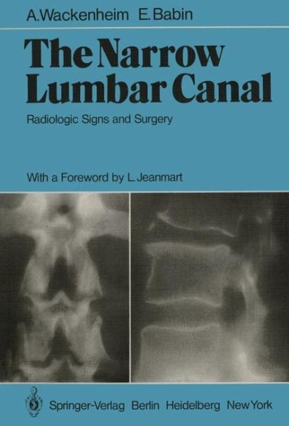
It is amazing to discover how little importance has been attached to narrow lumbar canal syndromes up to now. Though H. VERBIEST gave a very accurate description in 1949, the neurologist's and neurosurgeon's preoccupations were mainly focused on discal pathology, disregarding the problem of an exclusively bony origin in canalar stenosis. A. WACKENHEIM and E. BABIN have the merit of becoming aware of the impor tance and originality of this problem; they organized in the beautiful surround ings of the Bischenberg near Strasbourg, a postgraduate course, in which the most eminent European specialists in this field participated. I am very honored to have been asked to write the introduction to this mono graphy, which contains all the studies reported and commented on during this meeting. Before considering the problem from the various radiologic points of view, it is in my opinion indispensable to define the term "stenosis." We could not do so more accurately than by assuming the definition proposed by A. WACKENHEIM and E. BABIN and unanimously confirmed by all those who attented the session.Hinweis: Dieser Artikel kann nur an eine deutsche Lieferadresse ausgeliefert werden.
1. Radiology of the Narrow Lumbar Canal.- 1.1 History.- 1.2 Terminology.- 1.3 Anatomy.- 1.4 Clinical Data.- 1.5 Radiologic Techniques and Their Indications.- 1.6 Radiologic Signs of Lumbar Canal Narrowness.- 1.7 Nosology.- Figures 1-7.- 2. Plain X-Ray Diagnosis of Developmental Narrow Lumbar Canal.- 2.1 Technique.- 2.2 Findings: To Measure or Not to Measure? That is Not the Question.- 2.3 Requirements for Reliable Measurements and Pitfalls.- 2.4 Radiologic Features of the Narrow Lumbar Canal Without Contrast Medium.- 2.5 Findings on Lateral Projection.- 2.6 Various Types of Developmental Stenosis.- 2.7 Correlation Between Surgical and Radiologic Reports.- 2.8 Narrow Lumbar Canal and Associated Diseases.- Figures 8-14.- 3. Interapophysolaminar Spaces (IALS) of the Lumbar Spine and Their Utility in the Diagnosis of Narrow Lumbar Canal.- 3.1 Introduction.- 3.2 Material and Methods.- 3.3 Results.- 3.4 Conclusion.- Figures 15-18.- 4. Myelographic Signs of Narrow Lumbar Canal.- 4.1 Technical Particularities.- 4.2 Limits of the LM as an Investigation of the Narrow Lumbar Canals.- 4.3 LM Anomalies.- Figures 19-28.- 5. Gas Myelography in Verbiest's Developmental Spinal Canal Stenosis.- 5.1 Symptomatology.- 5.2 Radiologic Examination.- 5.3 Clinical Forms of Idiopathic Developmental Spinal Stenosis.- Figures 29-42.- 6. Phlebographic Signs of the Narrow Lumbar CanaL.- 6.1 Physiopathology of the Venous Compression.- 6.2 Phlebographic Signs of Narrow Lumbar Canal.- Figures 43-47.- 7. Narrow Lumbar Canal by Postoperative Epidural Lesions.- 7.1 Radiculosaccographic Semeiology of Epidural Scarring.- 7.2 Phlebographic Semeiology of Epidural Scarring.- 7.3 Surgical Findings.- 7.4 Clinical Aspects.- 7.5 Physiopathology.- 7.6 Conclusion.- Figures 48-60.- 8. SpinalPhlebography in the Stenosis of the Lumbar Canal.- Figures 61-72.- 9. Computerized Tomography in Lumbar Spinal Stenosis.- 9.1 Material and Methods.- 9.2 Results.- 9.3 Conclusion.- Figures 73-84.- 10. Lumbar Spinal Stenosis.- 10.1 Etiology.- 10.2 Symptomatology.- 10.3 Treatment.- Figures 85-98.- 11. Narrow Radicular Canal.- 11.1 Nosologic Importance of the Narrow Radicular Canal with Regard to the Narrow Lumbar Canal.- 11.2 Anatomy of the Radicular Canal.- 11.3 Etiologies.- 11.4 Symptomatology of the Narrow Radicular Canal.- 11.5 Radiologic Findings.- 11.6 Surgical Procedures.- 11.7 Conclusion.- Figures 99-108.- 12. Stenosis of the Bony Lumbar Vertebral Canal.- 12.1 Introduction.- 12.2 Historical Review: Evolution of the Idea.- 12.3 Nomenclature.- 12.4 Classification of the Types of Stenoses of the Lumbar Vertebral Canal.- 12.5 Semiological Aspects.- Figures 125-127.- 12.6 Surgical Treatment and Results.- 13. Cheirolumbar Dysostosis: Developmental Brachycheiry and Narrowness of the Lumbar Canal.- Figures 128-139.- References.- Author Index.
Es gelten unsere Allgemeinen Geschäftsbedingungen: www.buecher.de/agb
Impressum
www.buecher.de ist ein Internetauftritt der buecher.de internetstores GmbH
Geschäftsführung: Monica Sawhney | Roland Kölbl | Günter Hilger
Sitz der Gesellschaft: Batheyer Straße 115 - 117, 58099 Hagen
Postanschrift: Bürgermeister-Wegele-Str. 12, 86167 Augsburg
Amtsgericht Hagen HRB 13257
Steuernummer: 321/5800/1497