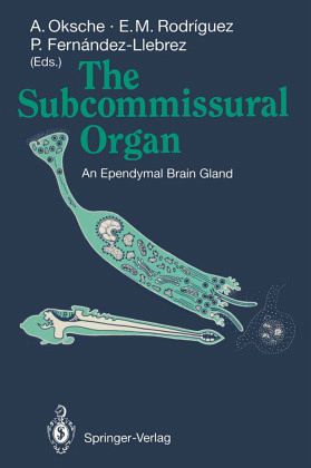
During the past two decades the progress in neuroendocrine research, stimulated by the increasing general interest in neurosciences, has been very impressive. Most of these efforts have concentrated on peuroendocrine nerve cells and their systems. Even if some aspects have remained open to discussion, the principal functional role of the neuroendocrine units capable of elaboration of biological active peptides (peptidergic neurons) is quite well understood. The same holds true for the central aminergic neurons and for such photoreceptor-derived paraneuronal elements as the pinealocytes. The…mehr
During the past two decades the progress in neuroendocrine research, stimulated by the increasing general interest in neurosciences, has been very impressive. Most of these efforts have concentrated on peuroendocrine nerve cells and their systems. Even if some aspects have remained open to discussion, the principal functional role of the neuroendocrine units capable of elaboration of biological active peptides (peptidergic neurons) is quite well understood. The same holds true for the central aminergic neurons and for such photoreceptor-derived paraneuronal elements as the pinealocytes. The primordium of the central nervous system possesses potencies for central sensory and secretory differentiations. Among the latter, a non-neuronal ependymal structure - the subcommissural orga- has remained enigmatic in terms of its biological significance. The sub commissural organ is a common, very constant, and conservative property of the vertebrate brain, from cyclostomes to mammals, and its appears early in ontogeny. The spectacular secretory activity of this brain gland, located in the diencephalic roof at the entrance to the mesencephalic aqueduct, results in the formation of an intraventricular secretory product - Reissner's fiber. This peculiar structural complex has attracted investigators to use a wide spectrum of modern cytological and, more recently, also molecular methods to investigate the secretory process and the secretory product, primarily glycoproteins, in greater detail. So far, however, the progress in structural insight has outpaced our knowledge of the function of the subcommissural organ.
I. Historical Reflections.- Ernst Reissner (1824-1878): Anatomist of the "Membrane" and "Fiber" (With 1 Figure).- Helmut Hofer (1912-1989) and the Concept of the Circumventricular Organs (With 1 Figure).- Historical Landmarks in the Investigation of the Subcommissural Organ and Reissner's Fiber (With 1 Figure).- II. Phylogeny and Ontogeny.- Phylogenetic and Conceptual Aspects of the Subcommissural Organ (With 3 Figures).- Reissner's Fiber Mechanisms: Some Common Denominators (With 3 Figures).- Ontogenetic Development of the Subcommissural Organ with Reference to the Flexural Organ (With 2 Figures).- Developmental Aspects of the Subcommissural Organ: An Approach Using Lectins and Monoclonal Antibodies (With 5 Figures).- The Subcommissural Organ and Ontogenetic Development of the Brain (With 5 Figures).- III. Biochemistry.- Protein Glycosylation in Mammalian Cells (With 3 Figures).- Partial Characterization of the Secretory Products of the Subcommissural Organ (With 3 Figures and 1 Table).- Biochemical and Immunochemical Analysis of Specific Compounds in the Subcommissural Organ of the Bovine and Chick (With 4 Figures and 3 Tables).- Immunochemical Analysis of the Dogfish Subcommissural Organ (With 2 Figures and 1 Table).- IV. Secretory Process, Including Model Systems.- Dynamic Aspects of the Secretory Process in the Amphibian Subcommissural Organ.- Evidence for the Release of CSF-Soluble Secretory Material from the Subcommissural Organ, with Particular Reference to the Situation in the Human (With 3 Figures).- The Subcommissural Organ In Vitro (With 2 Figures).- V. Relation to Other Circumventricular Organs: Conceptual Aspects.- Circumventricular Organs and Modulation in the Midsagittal Plane of the Brain (With 1 Figure and 1 Table).- Blood- andCerebrospinal Fluid-Dominated Compartments of the Rat Brain (With 1 Figure).- VI. Neural Inputs.- The Subcommissural Organ of the Rat: An In Vivo Model of Neuron-Glia Interactions (With 2 Figures and 1 Table).- Neural Inputs to the Subcommissural Organ (With 2 Figures).- Impairment of the Serotoninergic Innervation of the Rat Subcommissural Organ after Early Postnatal X-Irradiation of the Brain Stem (With 2 Figures).- Immunocytochemistry of Neuropeptides and Neuropeptide Receptors in the Subcommissural Organ of the Rat (With 10 Figures).- VII. Connections with Other Brain Structures.- Relationships Between the Subcommissural Organ and the Pineal Complex (With 2 Figures).- The Goldfish Subcommissural Organ Is Not Innervated by Fibers of the Pineal Tract (With 2 Figures).- Immunochemical Relationships Between the Subcommissural Organ and Hypothalamic Neurons (With 6 Figures and 1 Table).- VIII. Physiology of the Cerebrospinal Fluid.- Cerebrospinal Fluid Secretion: The Transport of Fluid and Electrolytes by the Choroid Plexus (With 4 Figures).- Physiology of Cerebrospinal Fluid Circulation: Amphibians, Mammals, and Hydrocephalus (With 6 Figures and 1 Table).- IX. Experimental Aspects.- Effects of Hibernation and Hypothermia on the Secretory Activity of the Subcommissural Organ (With 3 Figures).- The Subcommissural Organ: Immunohistochemistry and Potential Relations to Salt/Water Balance (With 6 Figures and 1 Table).- Immunological Blockade of the Subcommissural Organ - Reissner's Fiber Complex (With 6 Figures).- The Effect of Immunological Blockade of Reissner's Fiber Formation on the Circulation of Cerebrospinal Fluid Along the Central Canal of the Rat Spinal Cord (With 4 Figures).- X. Discussion Sessions (With 3 Figures).- I Signal Pathways.- II Cell Biology andBiochemistry.- III Functional Aspects I.- IV Functional Aspects II.
Es gelten unsere Allgemeinen Geschäftsbedingungen: www.buecher.de/agb
Impressum
www.buecher.de ist ein Internetauftritt der buecher.de internetstores GmbH
Geschäftsführung: Monica Sawhney | Roland Kölbl | Günter Hilger
Sitz der Gesellschaft: Batheyer Straße 115 - 117, 58099 Hagen
Postanschrift: Bürgermeister-Wegele-Str. 12, 86167 Augsburg
Amtsgericht Hagen HRB 13257
Steuernummer: 321/5800/1497