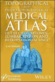
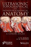
The fourth medical atlas in this new series on the human body and filled with detailed pictures, this atlas details the topographical and pathotopographical anatomy of the pelvis, spine, and limbs, a useful reference for medical professionals and students alike. Written by an experienced and well-respected physician and professor, this new volume, building on the previous volume, Ultrasonic Topographical and Pathotopographical Anatomy, and its sequels, also available from Wiley-Scrivener, presents the ultrasonic topographical and pathotopographical anatomy of the pelvis, spine, and limbs, offering further detail into these important areas for use by medical professionals. This series of atlases of topographic and pathotopographic human anatomy is a fundamental and practically important series designed for doctors of all specializations and students of medical schools. Here you can find almost everything that is connected with the topographic and pathotopographic human anatomy, including original graphs of logical structures of topographic anatomy and development of congenital abnormalities, topography of different areas in layers, pathotopography, computer and magnetic resonance imaging (MRI) of topographic and pathotopographic anatomy. Also you can find here new theoretical and practical sections of topographic anatomy developed by the author himself which are published for the first time. They are practically important for mastering the technique of operative interventions and denying possibility of iatrogenic complications during operations. This important new volume will be valuable to physicians, junior physicians, medical residents, lecturers in medicine, and medical students alike, either as a textbook or as a reference. It is a must-have for any physician's library. This groundbreaking new volume: * Using the same technology as the first two atlases by the same author, solves the problem of visualization of topographical and pathotopographical anatomy * Builds on the author's previous book, Ultrasonic Topographical and Pathotopographical Anatomy and the sequel, focusing solely on the areas of the pelvis, spine, and limbs * Goes beyond previous attempts, which are often drawings, by using state-of-the-art technology to show detailed physiological areas * Is for medical professionals of all types and levels, from students and professors to working doctors and other medical professionals doing research and development AUDIENCE: Physicians, interns, junior physicians, medical residents, medical students, specialists in surgery, traumatology, neurosurgery, oncology, urology, obstetrics and gynecology, dental surgery, anesthesiology, and other medical specialists concerned with different topographical and anatomical structures.
Hinweis: Dieser Artikel kann nur an eine deutsche Lieferadresse ausgeliefert werden.
Hinweis: Dieser Artikel kann nur an eine deutsche Lieferadresse ausgeliefert werden.

