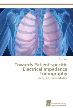
Mechanical ventilation of patients with severe lung injury is an important clinical treatment to ensure proper lung oxygenation and to mitigate the extent of collapsed lung regions. Electrical Impedance Tomography (EIT) is a novel method to determine functional processes inside the thorax such as lung ventilation and cardiac activity. Current EIT systems and algorithms use simplified or generalized thorax models to solve the reconstruction problem, which reduce image quality and anatomical significance. In this book, the development of a clinically relevant workflow to compute sophisticated three-dimensional thorax models from patient-specific CT data is described. The significantly improved image quality and anatomical precision of EIT images reconstructed with these 3D models is reported, and the impact on clinical applicability is discussed. The results presented in this book contribute significantly to clinical research efforts to pave the way towards improved patient-specific treatments of lung injury using EIT.
Hinweis: Dieser Artikel kann nur an eine deutsche Lieferadresse ausgeliefert werden.
Hinweis: Dieser Artikel kann nur an eine deutsche Lieferadresse ausgeliefert werden.

