40,95 €
40,95 €
inkl. MwSt.
Sofort per Download lieferbar
payback
20 °P sammeln
40,95 €
40,95 €
inkl. MwSt.
Sofort per Download lieferbar

Alle Infos zum eBook verschenken
payback
20 °P sammeln
Als Download kaufen
40,95 €
inkl. MwSt.
Sofort per Download lieferbar
payback
20 °P sammeln
Jetzt verschenken
40,95 €
inkl. MwSt.
Sofort per Download lieferbar

Alle Infos zum eBook verschenken
payback
20 °P sammeln
  • Format: PDF

This book introduces an exciting new method for breast ultrasound diagnostics - automated whole-breast volume scanning (3D ABVS). Scanning technique is described in detail, with guidance on scanning positions and protocols. Imaging findings are then illustrated and discussed for normal breast variants, the different forms of breast cancer, fibroadenomas, cystic disease, benign and malignant male breast disorders, mastitis, breast implants, and postoperative breast scars. In order to aid appreciation of the benefits of 3D ABVS, comparisons with findings on X-ray mammography and conventional 2D…mehr

Produktbeschreibung
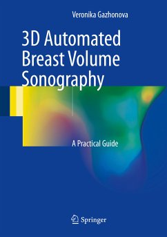
This book introduces an exciting new method for breast ultrasound diagnostics - automated whole-breast volume scanning (3D ABVS). Scanning technique is described in detail, with guidance on scanning positions and protocols. Imaging findings are then illustrated and discussed for normal breast variants, the different forms of breast cancer, fibroadenomas, cystic disease, benign and malignant male breast disorders, mastitis, breast implants, and postoperative breast scars. In order to aid appreciation of the benefits of 3D ABVS, comparisons with findings on X-ray mammography and conventional 2D hand-held US are presented. Readers will be especially impressed by the convincing demonstration of the advantages of the new method for diagnosis of breast cancer in women with dense glandular tissue. In enabling readers to learn how to perform and interpret 3D ABVS, this book will be of great value for all who are embarking on its use. It will also serve as a welcome reference for radiologists, oncologists, and ultrasonographers who already have some familiarity with the technique.


Dieser Download kann aus rechtlichen Gründen nur mit Rechnungsadresse in A, B, BG, CY, CZ, D, DK, EW, E, FIN, F, GR, HR, H, IRL, I, LT, L, LR, M, NL, PL, P, R, S, SLO, SK ausgeliefert werden.

Autorenporträt
Veronika Gazhonova, MD, PhD, is a consultant and chief ultrasound specialist at the United Hospital and Policlinic, Moscow, Russia and Professor of Radiology and Chief Radiology Chair in the Postgraduate Medical Education & Research Center, President Medical Center, Moscow. Her areas of clinical interest are innovations in breast ultrasonography, including 3D US, sonoelastography, US-guided procedures, and contrast US. She has previously published five books as well as more than 100 articles in Russian journals and 19 publications available via ResearchGate. Dr. Gazhonova is a member of the Russian Association of Radiology (RAR), the European Society of Radiology (since 1991), and the Russian Association of Ultrasound in Medicine and Biology (RAUMB).