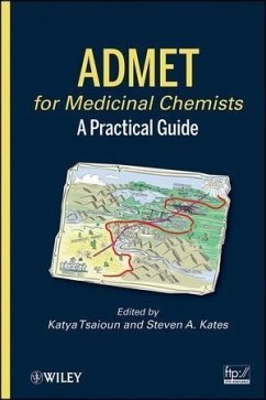
ADMET for Medicinal Chemists (eBook, ePUB)
A Practical Guide
Redaktion: Tsaioun, Katya; Kates, Steven A.
Alle Infos zum eBook verschenken
ADMET for Medicinal Chemists (eBook, ePUB)
A Practical Guide
Redaktion: Tsaioun, Katya; Kates, Steven A.
- Format: ePub
- Merkliste
- Auf die Merkliste
- Bewerten Bewerten
- Teilen
- Produkt teilen
- Produkterinnerung
- Produkterinnerung
Hier können Sie sich einloggen
Bitte loggen Sie sich zunächst in Ihr Kundenkonto ein oder registrieren Sie sich bei bücher.de, um das eBook-Abo tolino select nutzen zu können.
This book guides medicinal chemists in how to implement early ADMET testing in their workflow in order to improve both the speed and efficiency of their efforts. Although many pharmaceutical companies have dedicated groups directly interfacing with drug discovery, the scientific principles and strategies are practiced in a variety of different ways. This book answers the need to regularize the drug discovery interface; it defines and reviews the field of ADME for medicinal chemists. In addition, the scientific principles and the tools utilized by ADME scientists in a discovery setting, as…mehr
- Geräte: eReader
- mit Kopierschutz
- eBook Hilfe
- Größe: 4.72MB
Dieser Download kann aus rechtlichen Gründen nur mit Rechnungsadresse in A, B, BG, CY, CZ, D, DK, EW, E, FIN, F, GR, HR, H, IRL, I, LT, L, LR, M, NL, PL, P, R, S, SLO, SK ausgeliefert werden.
- Produktdetails
- Verlag: For Dummies
- Seitenzahl: 512
- Erscheinungstermin: 16. Februar 2011
- Englisch
- ISBN-13: 9780470922811
- Artikelnr.: 37300563
- Verlag: For Dummies
- Seitenzahl: 512
- Erscheinungstermin: 16. Februar 2011
- Englisch
- ISBN-13: 9780470922811
- Artikelnr.: 37300563
Contributors xix
1 Introduction 1
Corinne Kay
1.1 Introduction 1
1.2 Voyage Through The Digestive System 2
1.2.1 The Mouth 3
1.2.2 The Stomach 4
1.2.3 The Small Intestine: Duodenum 7
1.2.4 The Small and Large Intestine: Jejunum, Ileum, Colon 9
1.2.5 Hepatic-Portal Vein 13
1.3 The Liver Metabolism 15
1.3.1 CYP450 (CYPs) 17
1.4 The Kidneys 21
1.4.1 Active Tubular Secretion 23
1.4.2 Passive Tubular Reabsorption 24
1.5 Conclusions 25
References 25
2 In Silico ADME/Tox Predictions 29
David Lagorce, Christelle Reynes, Anne-Claude Camproux, Maria A. Miteva,
Olivier Sperandio, and Bruno O. Villoutreix
2.1 Introduction 29
2.2 Key Computer Methods for ADME/Tox Predictions 30
2.2.1 Drug Discovery 30
2.2.2 Applying or Not ADME/Tox Predictions, Divided Opinions 35
2.2.3 In Silico ADME/Tox Methods and Modeling Approaches 39
2.2.4 Physicochemistry, Pharmacokinetics, Drug-Like and Lead-Like Concepts
46
2.2.5 Lipophilicity 51
2.2.6 pKa 53
2.2.7 Transport Proteins 61
2.2.8 Plasma Protein Binding 62
2.2.9 Metabolism 65
2.2.10 Elimination 67
2.2.11 Toxicity 67
2.3 Preparation of Compound Collections and Computer Programs, Challenging
ADME/Tox Predictions and Statistical Methods 73
2.3.1 Preparation of Compound Collections and Computer Programs 73
2.3.2 Preparing a Compound Collection: Materials and Methods 75
2.3.3 Cleaning and Designing the Compound Collection 83
2.3.4 Searching for Similarity 85
2.3.5 Generating 3D Structures 86
2.4 ADME/Tox Predictions within Pharmaceutics Companies 86
2.4.1 Actelion Pharmaceuticals Ltd. 86
2.4.2 Bayer 86
2.4.3 Bristol-Myers Squibb 87
2.4.4 Hoffmann-La Roche Ltd. 87
2.4.5 Neurogen Corporation 87
2.4.6 Novartis 88
2.4.7 Schering AG 88
2.4.8 Vertex Pharmaceuticals 88
2.5 Challenging ADME/Tox Predictions 88
2.5.1 Tolcapone 89
2.5.2 Factor V Inhibitors 89
2.5.3 CRF-1 Receptor Antagonists 90
2.6 Statistical Methods 90
2.6.1 Principal Component Analysis 90
2.6.2 Partial Least Square 93
2.6.3 Support Vector Machine 96
2.6.4 Decision Trees 98
2.6.5 Neural Networks 101
2.7 Conclusions 104
References 105
3 Absorption and Physicochemical Properties of the NCE 125
Jon Selbo and Po-Chang Chiang
3.1. Introduction 125
3.2. Physicochemical Properties 126
3.3. Stability 127
3.4. Dissolution and Solubility 128
3.4.1. Dissolution Rate, Particle Size, and Solubility 128
3.4.2. pH and Salts 130
3.4.3. In Vivo Solubilization 133
3.5. Solid State 134
References 139
4 ADME 145
Martin E. Dowty, Dean M. Messing, Yurong Lai, and Leonid (Leo) Kirkovsky
4.1 Introduction 145
4.2 Absorption 146
4.2.1 Route of Administration 146
4.2.2 Factors Determining Oral Bioavailability 149
4.3 Distribution 157
4.3.1 Drug Distribution 157
4.3.2 Volume of Distribution 158
4.3.3 Free Drug Concentration 160
4.3.4 CNS Penetration 162
4.4 Elimination 165
4.4.1 Elimination Versus Clearance 165
4.4.2 Metabolism Versus Excretion 165
4.4.3 Drug-Free Fraction and Clearance 166
4.4.4 Lipophilicity and Clearance 166
4.4.5 Transporters and Clearance 166
4.4.6 Metabolism 167
4.4.7 Excretion 171
4.5 Drug Interactions 174
4.5.1 Absorption-Driven DDI 174
4.5.2 Distribution-Driven DDI 174
4.5.3 Excretion-Driven DDI 174
4.5.4 Metabolism-Driven DDI 175
4.5.5 Tools for Studying Drug Metabolism 177
4.5.6 Applications of Drug Metabolism Tools 180
4.5.7 Tools for Studying Drug Excretion 184
4.6 Strategies for Assessing ADME Properties 186
4.6.1 Assessing ADME Attributes at Different Stages of Discovery Projects
186
4.7 Tool Summary for Assessing ADME Properties 190
References 190
5 Pharmacokinetics for Medicinal Chemists 201
Leonid (Leo) Kirkovsky and Anup Zutshi
5.1 Introduction 201
5.1.1 History of Pharmacokinetics as Science 201
5.2 ADME 202
5.2.1 Absorption 202
5.2.2 Distribution 204
5.2.3 Metabolism 207
5.2.4 Excretion 207
5.3 The Mathematics of Pharmacokinetics 211
5.3.1 Compartmental Versus Noncompartmental Analysis 212
5.4 Drug Administration and PK Observations 212
5.4.1 Analysis of Intravenous PK Data 213
5.4.2 Analysis of Extravascular PK Data 227
5.4.3 Analysis of Intravenous Infusion Data 230
5.4.4 Analysis of PK Data after Multiple Dose Administrations 231
5.4.5 Analysis of PK Data after Escalating Dose Administrations 233
5.5 Human PK Projection 235
5.5.1 Allometric Scaling 235
5.5.2 Scaling by Physiologically Based Pharmacokinetic Modeling 237
5.5.3 In Vitro-In Vivo Correlations 239
5.6 PK Practices 239
5.6.1 PK Studies for Different Stages of Discovery Projects 240
5.6.2 Key Parameters of PK Studies 241
5.7 Engineering Molecules with Desired ADME Profile 269
5.A Appendices 269
5.A.1 General Morphinometric Data for Different Species 269
5.A.2 Organ Weights in Different Species 270
5.A.3 Organ, Tissue, and Fluid Volumes in Different Species 271
5.A.4 Blood Content in Different Rat Organs 271
5.A.5 Biofluid Flow through the Organs in Different Species 272
5.A.6 Anatomical Characteristics of GI Tract in Different Species 273
5.A.7 The pH and Motility of GI Tract in Different Species 274
5.A.8 Phase I and Phase II Metabolism in Different Species 274
Acknowledgments 277
References 277
6 Cardiac Toxicity 287
Ralf Kettenhofen and Silke Schwengberg
6.1 Introduction 287
6.2 Ion Channel-Related Cardiac Toxicity 287
6.2.1 Cardiac Electrophysiology 288
6.2.2 Delayed Repolarization: Mechanisms and Models 290
6.2.3 Shortened Ventricular Repolarization 294
6.2.4 Alterations in Intracellular Ca2þ Handling 296
6.2.5 Preclinical Models for Assessment of Ion Channel-Related
Cardiotoxicity 297
6.3 Nonarrhythmic Cardiac Toxicity 299
6.3.1 Definition of Drug-Induced Cardiac Toxicity 300
6.3.2 Assays for Detection of Nonarrhythmic Cardiac Toxicity 300
6.3.3 Biochemical and Molecular Basis of Drug-Induced Cardiac
Toxicity-Impairment of Mitochondrial Function 304
References 306
7 Genetic Toxicity: In Vitro Approaches for Medicinal Chemists 315
Richard M. Walmsley and David Elder
7.1 Introduction 315
7.1.1 Scope of this Chapter 315
7.1.2 Definitions 316
7.1.3 Positive Genotoxicity Data is not Uncommon and Very Costly 316
7.1.4 Why Genome Damage is Undesirable 317
7.1.5 The Inherent Integrity of the Genome and its Inevitable Corruption
317
7.1.6 Many Chemicals can Cause Cancer, but do not Pose a Significant Risk
to Humans 318
7.1.7 The False Positives: Many Chemicals Produce Positive Genotoxicity
Data that are neither Carcinogens nor In Vivo Genotoxins 318
7.1.8 Defense Against Genotoxic Damage 319
7.1.9 Mechanisms of Genotoxic Damage 320
7.1.10 Genotoxicity Assessment Occurs after Medicinal Chemistry
Optimization 321
7.2 Limitations in the Regulatory In Vitro Genotoxicity Tests 322
7.2.1 Biology Limitations of In Vitro Tests 322
7.2.2 Hazard and Safety Assessment have Different Requirements 323
7.2.3 The Data from Genetic Toxicologists 323
7.3 Practical Issues for Genotoxicity Profiling 324
7.3.1 Vehicle 324
7.3.2 Dilution Range 324
7.3.3 Purity 324
7.4 Computational Approaches to Genotoxicity Assessment: The In Silico
Methods 325
7.4.1 General Considerations 325
7.4.2 The Chemistry of Genotoxins 328
7.5 Genotoxicity Assays for Screening 335
7.5.1 Bacterial Gene Mutation Assays 337
7.5.2 Mammalian Cell Mutation Assays 338
7.5.4 Chromosome Damage and Aberration Assays 339
7.5.5 The .Comet. Assay 340
7.5.6 DNA Adduct Assessment 341
7.5.7 Gene Expression Assays 341
7.6 The .Omics. 343
7.7 Using Data from In Vitro Profiling: Confirmatory Tests, Follow-Up
Tests, and the Link to Safety Assessment and In Vivo Models 343
7.7.1 Annotations from Screening Data 344
7.7.2 Can a Genetic Toxicity Profile Assist with In Vivo Testing
Strategies? 344
7.8 What to Test, When, and How 345
7.9 Changes to Regulatory Guidelines Can Influence Screening Strategy 346
7.10 Summary 347
Acknowledgment 347
References 348
8 Hepatic Toxicity 353
Jinghai James Xu and Keith Hoffmaster
8.1 Introduction 353
8.2 Mechanisms of DILI 354
8.2.1 Reactive Metabolite Formation 355
8.2.2 Mitochondrial Dysfunction and Oxidative Stress 357
8.2.3 Bile Flow, Drug-Induced Cholestasis, and Inhibition of Biliary Efflux
Transporters 359
8.3 Assays and Test Systems to Measure Various Types of DILI 360
8.4 Medicinal Chemistry Strategies to Minimize DILI 365
8.5 Future Outlooks 370
Acknowledgment 370
References 370
9 In Vivo Toxicological Considerations 379
John P. Devine, Jr.
9.1 Introduction 379
9.2 Route of Administration 379
9.2.1 Oral Route 380
9.2.2 Intravenous Route 381
9.2.3 Dermal Route 382
9.3 Formulation Issues 383
9.4 Compound Requirements 384
9.5 Animal Models 385
9.5.1 Mouse 385
9.5.2 Rat 386
9.5.3 Dog 386
9.5.4 Swine 386
9.5.5 Nonhuman Primates 387
9.6 IND-Supporting Toxicology Studies 387
9.6.1 Single-Dose Studies 387
9.6.2 Repeat-Dose Studies 388
9.7 Study Result Interpretation 392
9.7.1 Clinical Observations 392
9.7.2 Body Weight/Feed Consumption 393
9.7.3 Clinical Pathology 393
9.7.4 Clinical Chemistry 393
9.7.5 Electrocardiograms 394
9.7.6 Organ Weights 394
9.7.7 Pathology 395
9.8 Genetic Toxicology Studies 395
9.8.1 Gene Mutation 395
9.8.2 Chromosomal Aberration 396
9.8.3 In Vivo Mouse Micronucleus 396
9.9 Conclusion 396
References 397
10 Preclinical Candidate Nomination and Development 399
Nils Bergenhem
10.1 Introduction 399
10.2 Investigational New Drug Application and Clinical Development 400
10.2.1 Chemistry, Manufacturing, and Control Information 401
10.2.2 Animal Pharmacology and Toxicology Studies 401
10.2.3 Clinical Protocols and Investigator Information 401
10.3 Strategic Goals for the Preclinical Development 402
10.4 Selection of Preclinical Development Candidate 403
10.4.1 Efficacy 403
10.4.2 Safety/Tolerance 405
10.4.3 PK 407
10.4.4 Non-GLP Toxicological Study 407
10.5 CMC 408
10.5.1 Solubility 408
10.5.2 Solutions Stability 408
10.5.3 Synthetic Feasibility, Solid-State Stability, and Hygroscopicity 408
10.5.4 Patent Position 408
10.6 Preclinical Studies 409
10.6.1 Example 1: IND Enabling Data Package to Support 1 Month Dosing in
Man 410
10.6.2 Example 2: Peroxisome Proliferator-Activated Receptor Agonist for
Type-2 Diabetes 410
10.6.3 Mass Balance 410
10.6.4 Animal Pharmacology and Toxicology Studies 410
10.6.5 Regulatory 414
10.7 Conclusions 415
References 415
11 Fragment-Based Drug Design: Considerations for Good ADME Properties 417
Haitao Ji
11.1 Introduction 417
11.2 Fragment-Based Screening 418
11.2.1 Fragment Library Design 419
11.2.2 Detection and Characterization of Weakly Binding Ligands 420
11.2.3 Approaches from Fragment to Lead Structures 427
11.3 Case Studies of Fragment-Based Screening for Better Bioavailability
431
11.3.1 Adenosine Kinase 431
11.3.2 Leukocyte Function-Associated Antigen-1 432
11.3.3 Matrix Metalloproteinase 3 (Stromelysins) 432
11.3.4 Protein Tyrosine Phosphatase 1B 433
11.3.5 b-Secretase (BACE-1) 436
11.3.6 SH2 Domain of pp60Src [62, 129] 439
11.3.7 Thrombin 439
11.3.8 Urokinase 441
11.3.9 Cathepsin S 442
11.3.10 Caspase-3 442
11.3.11 HIV-1 Protease 444
11.4 De Novo Design 445
11.4.1 In Silico Fragment Screening 447
11.4.2 Scaffold Hopping 448
11.5 Case Studies of De Novo Design for Better Bioavailability 450
11.5.1 DNA Gyrase 450
11.5.2 Factor Xa 450
11.5.3 X-Linked Inhibitor of Apoptosis Protein 451
11.5.4 Activator Protein-1 [196b] 451
11.6 Minimal Pharmacophoric Elements and Fragment Hopping 452
11.6.1 Minimal Pharmacophoric Elements 452
11.6.2 Fragment Hopping 453
11.6.3 Case Study: Nitric Oxide Synthase 457
11.7 Conclusions and Future Perspectives 459
Acknowledgments 460
References 460
Index 487