96,95 €
96,95 €
inkl. MwSt.
Sofort per Download lieferbar
payback
48 °P sammeln
96,95 €
96,95 €
inkl. MwSt.
Sofort per Download lieferbar

Alle Infos zum eBook verschenken
payback
48 °P sammeln
Als Download kaufen
96,95 €
inkl. MwSt.
Sofort per Download lieferbar
payback
48 °P sammeln
Jetzt verschenken
96,95 €
inkl. MwSt.
Sofort per Download lieferbar

Alle Infos zum eBook verschenken
payback
48 °P sammeln
  • Format: ePub

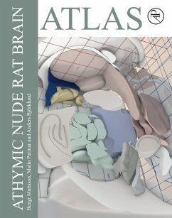
Athymic Nude Rat Brain Atlas will provide the first stereotaxic brain atlas of the athymic nude rat (Crl:NIH-Foxn1rnu), a T-cell deficient rat model commonly used in experimental studies and pre-clinical safety and efficacy studies. This 2D vector-based atlas contains coronal, sagittal, and horizontal brain sections of an athymic rat brain rendered from a single cleared specimen, placed in a computerized 3D environment. The maps enable readers to better calculate co-ordinates to target specific structures for toxin, virus, or cell delivery using stereotaxic surgery. This atlas will be a…mehr

Produktbeschreibung
Athymic Nude Rat Brain Atlas will provide the first stereotaxic brain atlas of the athymic nude rat (Crl:NIH-Foxn1rnu), a T-cell deficient rat model commonly used in experimental studies and pre-clinical safety and efficacy studies. This 2D vector-based atlas contains coronal, sagittal, and horizontal brain sections of an athymic rat brain rendered from a single cleared specimen, placed in a computerized 3D environment. The maps enable readers to better calculate co-ordinates to target specific structures for toxin, virus, or cell delivery using stereotaxic surgery. This atlas will be a valuable resource for any neuroscientist who wishes to work with nude rats in experimental and pre-clinical studies.

  • Contains coronal, sagittal, and horizontal maps of young adult athymic nude rat brain, spaced with a distance of 0.2 or 0.25 mm
  • Uses "flat skull" Bregma and Lambda as anatomical landmarks for correct placement in the 3D environment
  • Anatomical structures and nomenclature follow the standard set by the Paxinos and Watson rat brain atlases
  • Includes a map of the dopamine projection system as well as the distribution of the A8-A14 dopamine cell groups
  • Allows for easy read-out of coordinates for precise injections using stereotactic surgery

Dieser Download kann aus rechtlichen Gründen nur mit Rechnungsadresse in A, B, BG, CY, CZ, D, DK, EW, E, FIN, F, GR, HR, H, IRL, I, LT, L, LR, M, NL, PL, P, R, S, SLO, SK ausgeliefert werden.


Hinweis: Dieser Artikel kann nur an eine deutsche Lieferadresse ausgeliefert werden.
Autorenporträt
Mattsson is a Technician in Developmental and Regenerative Neurobiology within the Department of Experimental Medical Science at Wallenberg Neuroscience Center in Lund¿University. His research focus is on multidisciplinary research in Parkinson's Disease to create improved and novel treatments, disease modifications, and eventually cures to improve the quality of life for people living and ageing with these disorders.Dr. Parmar PhD is Professor of Developmental and Regenerative Neurobiology at Lund University. She is a New York Stem Cell Foundation Robertson Investigator. She was awarded the Bengt Falck Prize in Neuroscience in 2018 for pioneering stem cell research that has opened new possibilities for treating patients with neurodegenerative diseases, and also the Eric K. Fernström Prize in 2016 for young, promising, and successful researchers. The focus of her research is to understand cell fate specification in the developing brain and in human neural progenitor cells using cell-based models of neuronal differentiation - with the ultimate aim of developing these cells and technologies for use in brain repair.Dr. Björklund PhD is Senior Professor of Developmental and Regenerative Neurobiology and head of the Neurobiology unit at the Wallenberg Neuroscience Center at Lund University. He is a recently elected member of the National Academy of Sciences, is currently a professor. In 2011, he received the Robert A. Pritzker Prize for Leadership in Parkinson Research from the Michael J. Fox Foundation. His research group pioneered studies of neural transplantation to the brain in the 1970s, and the Lund neural transplantation program he leads has been one of the leading clinical programs for the development of restorative therapies in Parkinson's disease worldwide. During the last decade his lab has pioneered the use of the viral vectors for neurotrophic factor delivery to the brain and has been in the forefront of the development of this technology as a neuroprotective and restorative factor for the nigrostriatal dopamine system and its application in Parkinson's disease.