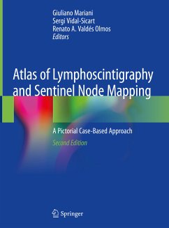
Atlas of Lymphoscintigraphy and Sentinel Node Mapping (eBook, PDF)
A Pictorial Case-Based Approach
Redaktion: Mariani, Giuliano; Valdés Olmos, Renato A.; Manca, Gianpiero
Versandkostenfrei!
Sofort per Download lieferbar
128,95 €
inkl. MwSt.

PAYBACK Punkte
64 °P sammeln!
Although lymphoscintigraphy was originally introduced into clinical routine for identification of the cause of peripheral edema, more recently it has been widely applied for radioguided biopsy of the sentinel lymph node in patients with solid cancers. The procedure is now considered crucial for adequate planning of oncologic surgery in a growing number of cancers. This atlas presents a collection of richly illustrated teaching cases that demonstrate the clinical relevance and impact of lymphoscintigraphy in different pathologic conditions. After introductory chapters on the anatomy, physiology...
Although lymphoscintigraphy was originally introduced into clinical routine for identification of the cause of peripheral edema, more recently it has been widely applied for radioguided biopsy of the sentinel lymph node in patients with solid cancers. The procedure is now considered crucial for adequate planning of oncologic surgery in a growing number of cancers. This atlas presents a collection of richly illustrated teaching cases that demonstrate the clinical relevance and impact of lymphoscintigraphy in different pathologic conditions. After introductory chapters on the anatomy, physiology, and pathophysiology of lymphatic circulation, the role of lymphoscintigraphy in differential diagnosis of peripheral edema and characterization of intracavitary lymph effusions is addressed. The principal focus of the book, however, is on the use of lymphoscintigraphic mapping for radioguided sentinel node biopsy in cutaneous melanoma and cancers at a range of anatomic sites. The most commonly observed lymphoscintigraphic patterns are depicted, and anatomic variants and technical pitfalls of the procedure receive careful attention. The role of tomographic multimodality imaging is also considered. The atlas will be an excellent learning tool for residents in nuclear medicine and other specialists with an interest in the field.
Dieser Download kann aus rechtlichen Gründen nur mit Rechnungsadresse in A, B, BG, CY, CZ, D, DK, EW, E, FIN, F, GR, HR, H, IRL, I, LT, L, LR, M, NL, PL, P, R, S, SLO, SK ausgeliefert werden.


