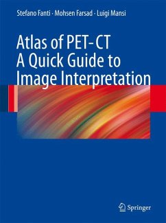
This user-friendly atlas, featuring about 500 images, should be a quick guide to interpreting PET/CT images with FDG in oncology. It also illustrates how to recognize normal, para-physiological, and benign pathological uptakes in a case-based practical manner. The text, which includes most relevant technical and pathophysiological premises, covers the main clinical applications and clearly articulates learning points and pitfalls. This atlas aims to become a standard text for nuclear medicine physicians and radiologists, residents and technicians whose work involves PET/CT imaging. This book is also suitable for both undergraduate and postgraduate courses.
Dieser Download kann aus rechtlichen Gründen nur mit Rechnungsadresse in A, B, BG, CY, CZ, D, DK, EW, E, FIN, F, GR, HR, H, IRL, I, LT, L, LR, M, NL, PL, P, R, S, SLO, SK ausgeliefert werden.

