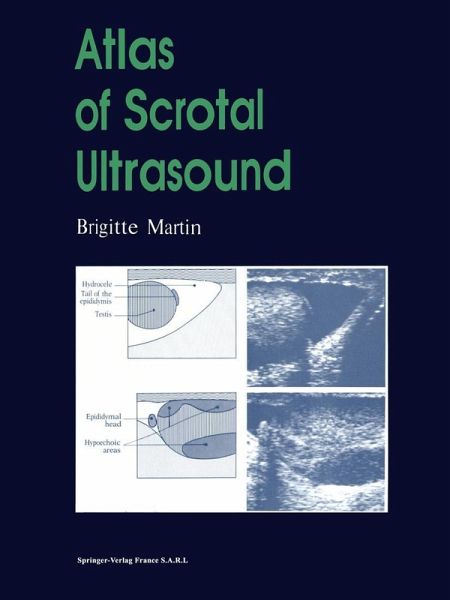
Atlas of Scrotal Ultrasound (eBook, PDF)
Versandkostenfrei!
Sofort per Download lieferbar
40,95 €
inkl. MwSt.
Weitere Ausgaben:

PAYBACK Punkte
20 °P sammeln!
The Atlas of Scrotal Ultrasound covers all the pathology related to the scrotal contents in the form of chapters corresponding to the main clinical presentations encountered in clinical practice; the acute scrotum, the chronically enlarged scrotum, the post-operative scrotum, infertility, cysts, calcifications. There are over 710 illustrations, each accompanied by a labelled diagram and description.
Dieser Download kann aus rechtlichen Gründen nur mit Rechnungsadresse in A, B, BG, CY, CZ, D, DK, EW, E, FIN, F, GR, HR, H, IRL, I, LT, L, LR, M, NL, PL, P, R, S, SLO, SK ausgeliefert werden.


