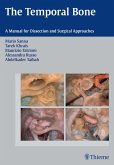
A very good knowledge of anatomical sections is necessary to be able to interpret fine imaging of the temporal bone. Both serial histological sections and CT material are presented in this volume. Three planes of space - horizontal, frontal and sagittal - are presented. The very thin histological slices make it possible to identify and study the vascular and nervous structures, section by section in and around the temporal bone. This very fine reading of the anatomical slices is an ideal aid for the clinician who must interpret normal and pathological CT sections.
Dieser Download kann aus rechtlichen Gründen nur mit Rechnungsadresse in A, B, BG, CY, CZ, D, DK, EW, E, FIN, F, GR, HR, H, IRL, I, LT, L, LR, M, NL, PL, P, R, S, SLO, SK ausgeliefert werden.


