Sie sind bereits eingeloggt. Klicken Sie auf 2. tolino select Abo, um fortzufahren.
Bitte loggen Sie sich zunächst in Ihr Kundenkonto ein oder registrieren Sie sich bei bücher.de, um das eBook-Abo tolino select nutzen zu können.
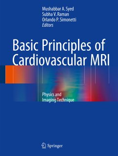
This book is a comprehensive and authoritative text on the expanding scope of CMR, dedicated to covering basic principles in detail focusing on the needs of cardiovascular imagers. The target audience for this book includes CMR specialists, trainees in CMR and cardiovascular medicine, cardiovascular physicists or clinical cardiovascular imagers. This book includes figures and CMR examples in the form of high-resolution still images and is divided in two sections: basic MRI physics, i.e. the nuts and bolts of MR imaging; and imaging techniques (pulse sequences) used in cardiovascular MR…mehr
This book is a comprehensive and authoritative text on the expanding scope of CMR, dedicated to covering basic principles in detail focusing on the needs of cardiovascular imagers. The target audience for this book includes CMR specialists, trainees in CMR and cardiovascular medicine, cardiovascular physicists or clinical cardiovascular imagers. This book includes figures and CMR examples in the form of high-resolution still images and is divided in two sections: basic MRI physics, i.e. the nuts and bolts of MR imaging; and imaging techniques (pulse sequences) used in cardiovascular MR imaging. Each imaging technique is discussed in a separate chapter that includes the physics and clinical applications (with cardiovascular examples) of a particular technique. Evolving techniques or research based techniques are discussed as well. This section covers both cardiac and vascular imaging. Cardiovascular magnetic resonance (CMR) imaging is now considered a clinically important imaging modality for patients with a wide variety of cardiovascular diseases. Recent developments in scanner hardware, imaging sequences, and analysis software have led to 3-dimensional, high-resolution imaging of the cardiovascular system. These developments have also influenced a wide variety of cardiovascular imaging applications and it is now routinely used in clinical practice in CMR laboratories around the world. The non-invasiveness and lack of ionizing radiation exposure make CMR uniquely important for patients whose clinical condition requires serial imaging follow-up. This is particularly true for patients with congenital heart disease (CHD) with or without surgical corrections who require lifelong clinical and imaging follow-up.
Dr. Mushabbar Syed is an associate professor of medicine and radiology at Stritch School of Medicine, Loyola University of Chicago USA. He also serves as the director of cardiovascular imaging. Prior to his current position, Dr. Syed was an associate professor of medicine and radiology and director of cardiovascular CT & MRI imaging at University of Kentucky, Lexington, Kentucky and assistant professor of medicine and radiology at Emory University School of Medicine, Atlanta, Georgia. Dr. Syed completed his medicine and cardiology training at Henry Ford Hospital, Detroit, Michigan followed by a clinical/research fellowship in cardiovascular MRI at the National Heart, Lung & Blood Institute, National Institutes of Health, Bethesda, Maryland. He has recently co-edited a textbook titled “Magnetic Resonance Imaging in Congenital Heart Disease” published by Springer Inc. 2012. Subha Raman, MD, MSEE, FACC, is a cardiologist and associate professor of medicine at The Ohio State University Medical Center. After undergraduate and graduate studies in electrical engineering as a National Science Foundation Fellow, Dr. Raman completed medical training including fellowship in cardiovascular medicine at The Ohio State University College of Medicine, with additional training in cardiovascular magnetic resonance (CMR) at Wake Forest University. Dr. Raman has published more than 70 papers and was named one of America’s Best Doctors in 2009. Her clinical interests include family risk and heritable heart disease, women’s cardiovascular health, early atherosclerosis detection and management, and improved stress testing. Dr. Raman’s research team works to develop innovations in cardiovascular imaging that provide earlier detection, more accurate diagnosis and personalized treatment of heart and vascular disease.
Inhaltsangabe
MRI scanner and coils.- Signal generation.- k-space.- Pulse sequence.- Image acquisition.- Image resolution.- Fast imaging.- High field MRI (3 Tesla or higher) .- Artifacts.- MRI safety.- Gating.- Gradient echo and cine imaging.- Black blood imaging and tissue characterization.- Perfusion.- Stress testing.- Late gadolinium enhancement imaging (scar/fibrosis) .- Phase contrast flow.- Coronary imaging.- Spectroscopy.- Contrast media.- Contrast enhanced MRA.- Non-contrast enhanced MRA.- Advanced cardiovascular magnetic resonance techniques.
Rezensionen
"The text is aimed at fellowship trainees but also as an aide-memoire for the established practitioner. It would be of great assistance to cardiovascular radiographers, explaining clearly many of the artefacts associated with this complex imaging technique and providing practical solutions. ... In a world where the time to understand and develop new techniques and services becomes fewer, as the pressure to deliver scans or scan reports increases, this book is an essential addition to an individual's or department's library." (Dr. Roger Bury, RAD Magazine, December, 2016)
Es gelten unsere Allgemeinen Geschäftsbedingungen: www.buecher.de/agb
Impressum
www.buecher.de ist ein Internetauftritt der buecher.de internetstores GmbH
Geschäftsführung: Monica Sawhney | Roland Kölbl | Günter Hilger
Sitz der Gesellschaft: Batheyer Straße 115 - 117, 58099 Hagen
Postanschrift: Bürgermeister-Wegele-Str. 12, 86167 Augsburg
Amtsgericht Hagen HRB 13257
Steuernummer: 321/5800/1497