73,95 €
73,95 €
inkl. MwSt.
Sofort per Download lieferbar
payback
37 °P sammeln
73,95 €
73,95 €
inkl. MwSt.
Sofort per Download lieferbar

Alle Infos zum eBook verschenken
payback
37 °P sammeln
Als Download kaufen
73,95 €
inkl. MwSt.
Sofort per Download lieferbar
payback
37 °P sammeln
Jetzt verschenken
73,95 €
inkl. MwSt.
Sofort per Download lieferbar

Alle Infos zum eBook verschenken
payback
37 °P sammeln
  • Format: PDF

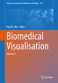
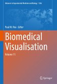
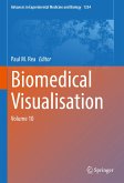
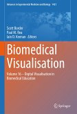
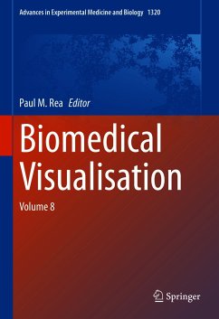
This edited book explores the use of technology to enable us to visualise the life sciences in a more meaningful and engaging way. It will enable those interested in visualisation techniques to gain a better understanding of the applications that can be used in visualisation, imaging and analysis, education, engagement and training. The reader will be able to explore the utilisation of technologies from a number of fields to enable an engaging and meaningful visual representation of the biomedical sciences, with a focus in this volume related to anatomy, and clinically applied scenarios. The…mehr

Produktbeschreibung
This edited book explores the use of technology to enable us to visualise the life sciences in a more meaningful and engaging way. It will enable those interested in visualisation techniques to gain a better understanding of the applications that can be used in visualisation, imaging and analysis, education, engagement and training.
The reader will be able to explore the utilisation of technologies from a number of fields to enable an engaging and meaningful visual representation of the biomedical sciences, with a focus in this volume related to anatomy, and clinically applied scenarios.
The first six chapters in this volume show the wide variety of tools and methodologies that digital technologies and visualisation techniques can be utilised and adopted in the educational setting. This ranges from body painting, clinical neuroanatomy, histology and veterinary anatomy through to real time visualisations and the uses of digital and social media for anatomical education. The last four chapters represent the diversity that technology has to be able to use differing realities and 3D capture in medical visualisation, and how remote visualisation techniques have developed. Finally, it concludes with an analysis of image overlays and augmented reality and what the wider literature says about this rapidly evolving field.

Dieser Download kann aus rechtlichen Gründen nur mit Rechnungsadresse in A, B, BG, CY, CZ, D, DK, EW, E, FIN, F, GR, HR, H, IRL, I, LT, L, LR, M, NL, PL, P, R, S, SLO, SK ausgeliefert werden.

Autorenporträt
Paul is a Professor of Digital and Anatomical Education at the University of Glasgow. He is qualified with a medical degree (MBChB), a MSc (by research) in craniofacial anatomy/surgery, a PhD in neuroscience, the Diploma in Forensic Medical Science (DipFMS), and an MEd with Merit (Learning and Teaching in Higher Education). He is an elected Fellow of the Royal Society for the encouragement of Arts, Manufactures and Commerce (FRSA), elected Fellow of the Royal Society of Biology (FRSB), Senior Fellow of the Higher Education Academy, professional member of the Institute of Medical Illustrators (MIMI) and a registered medical illustrator with the Academy for Healthcare Science. Paul has published widely and presented at many national and international meetings, including invited talks. He sits on the Executive Editorial Committee for the Journal of Visual Communication in Medicine, is Associate Editor for the European Journal of Anatomy and reviews for 25 different journals/publishers. He is the Public Engagement and Outreach lead for anatomy coordinating collaborative projects with the Glasgow Science Centre, NHS and Royal College of Physicians and Surgeons of Glasgow. Paul is also a STEM ambassador and has visited numerous schools to undertake outreach work. His research involves a long-standing strategic partnership with the School of Simulation and Visualisation The Glasgow School of Art. This has led to multi-million pound investment in creating world leading 3D digital datasets to be used in undergraduate and postgraduate teaching to enhance learning and assessment. This successful collaboration resulted in the creation of the worlds first taught MSc Medical Visualisation and Human Anatomy combining anatomy and digital technologies. The Institute of Medical Illustrators also accredits it. It has created college-wide, industry, multi-institutional and NHS research linked projects for students. Paul is the Programme Director forthis degree.