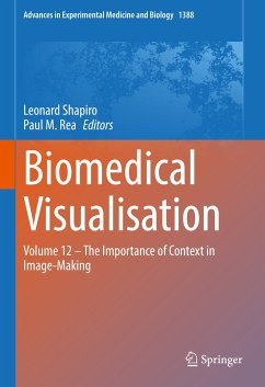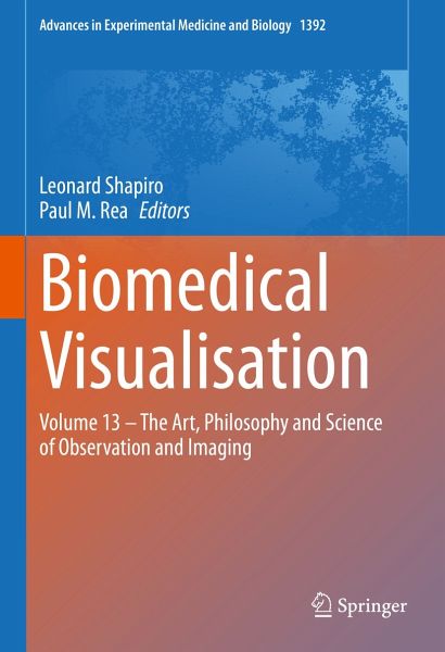
Sie sind bereits eingeloggt. Klicken Sie auf 2. tolino select Abo, um fortzufahren.
Bitte loggen Sie sich zunächst in Ihr Kundenkonto ein oder registrieren Sie sich bei bücher.de, um das eBook-Abo tolino select nutzen zu können.
This book brings together current advances in high-technology visualisation and the age-old but science-adapted practice of drawing for improved observation in medical education and surgical planning and practice. We begin this book with a chapter reviewing the history of confusion around visualisation, observation and theory, outlining the implications for medical imaging. The authors consider the shifting influence of various schools of philosophy, and the changing agency of technology over time. We then follow with chapters on the practical application of visualisation and observation,…mehr
This book brings together current advances in high-technology visualisation and the age-old but science-adapted practice of drawing for improved observation in medical education and surgical planning and practice.
We begin this book with a chapter reviewing the history of confusion around visualisation, observation and theory, outlining the implications for medical imaging. The authors consider the shifting influence of various schools of philosophy, and the changing agency of technology over time. We then follow with chapters on the practical application of visualisation and observation, including emerging imaging techniques in anatomy for teaching, research and clinical practice - innovation in the mapping of orthopaedic fractures for optimal orthopaedic surgical guidance - placental morphology and morphometry as a prerequisite for future pathological investigations - visualising the dural venous sinuses using volume tracing.
Two chapters explore the use and benefit of drawing in medical education and surgical planning. It is worth noting that experienced surgeons and artists employ a common set of techniques as part of their work which involves both close observation and the development of fine motor skills and sensitive tool use.
An in-depth look at police identikit construction from memory by eyewitnesses to crimes, outlines how an individual’s memory of a suspect’s facial features are rendered visible as a composite image.
This book offers anatomy educators and clinicians an overview of the history and philosophy of medical observation and imaging, as well as an overview of contemporary imaging technologies for anatomy education and clinical practice. In addition, we offer anatomy educators and clinicians a detailed overview of drawing practices for the improvement of anatomical observation and surgical planning. Forensic psychologists and law enforcement personnel will not only benefit from a chapter dedicated to the construction of facial composites, but also from chapters on drawing and observation.
Leonard is an artist working in the field of anatomy education with the Department of Human Biology at the University of Cape Town, South Africa. He has developed a novel, multi-sensory observation method that specifically employs the sense of touch (haptics) coupled with the simultaneous act of drawing. It is called the Haptico-visual observation and drawing (HVOD) method.
In anatomy education, the benefits of observing using the HVOD method include the enhanced observation of the three-dimensional (3D) form of different anatomical parts of the human body, the memorisation of these parts as a 3D 'mental picture', improved 3D spatial awareness, and an ability to draw. HVOD is taught in group workshops online or in physical classroom settings and is offered to medical students and clinicians. Leonard has taught the HVOD method at the University of Cape Town (South Africa), Newcastle University (England), The University of British Columbia (Canada), Carnegie Mellon University (USA). University College Cork (Ireland), Weill Cornell Medical College (USA), The Gordon Museum of Pathology at King’s College London (England), University of Texas Southwestern Medical School (USA). Leonard contributes to the anatomy education discourse via publications and articles, as an invited speaker and by presenting at anatomy conferences. Leonard Shapiro BSocSc, BA Fine Art (Hons), University of Cape Town.
Paul M. Rea
Paul is Professor of Digital and Anatomical Education at the University of Glasgow. He is Director of Innovation, Engagement and Enterprise within the School of Medicine, Dentistry and Nursing. He is also a Senate Assessor for Student Conduct, Council Member on Senate and coordinates the day-to-day running of the Body Donor Program and is a Licensed Teacher of Anatomy, licensed by the Scottish Parliament.
He is qualified with a medical degree (MBChB), a MSc (by research)in craniofacial anatomy/surgery, a PhD in neuroscience, the Diploma in Forensic Medical Science (DipFMS), and an MEd with Merit (Learning and Teaching in Higher Education). He is a senior fellow of the Higher Education Academy, a Fellow of the Institute of Medical Illustrators (FIMI) and a registered medical illustrator with the Academy for Healthcare Science.
Paul has published widely and presented at many national and international meetings, including invited talks. He has been the lead Editor for Biomedical Visualisation over 12 published volumes and is the founding editor for this book series. This has resulted in almost 85,000 downloads across these volumes, with contributions from over 450 different authors, across approximately 100 institutions from 19 countries across the globe. He sits on the Executive Editorial Committee for the Journal of Visual Communication in Medicine, is Associate Editor for the European Journal of Anatomy and has reviewed for 25 different journals/publishers.
He is the Public Engagement and Outreach lead for anatomy coordinating collaborative projects with the Glasgow Science Centre, NHS and Royal College of Physicians and Surgeons of Glasgow. Paul is also a STEM ambassador and has visited numerous schools to undertake outreach work.
His research involves a long-standing strategic partnership with the School of Simulation and Visualisation of the Glasgow School of Art. This has led to multi-million-pound investment in creating world-leading 3D digital datasets to be used in undergraduate and postgraduate teaching to enhance learning and assessment. This successful collaboration resulted in the creation of the world’s first taught MSc Medical Visualisation and Human Anatomy combining anatomy and digital technologies. The Institute of Medical Illustrators also accredits it. It has created college-wide, industry, multi-institutional and NHS research linked projects for students.
Inhaltsangabe
Part I. The History and Philosophy of Medical Imaging.- Chapter 1. Re:Viewing Observation. The Philosophy of Medical Imaging.- Part II. Innovation in Imaging Technologies.- Chapter 2. Emerging Imaging Techniques in Anatomy: For Teaching, Research and Clinical Practice.- Chapter 3. Mapping of Orthopaedic Fractures for Optimal Surgical Guidance.- Chapter 4. Visualising the Dural Venous Sinuses Using Volume Tracing, a Novel Approach.- Chapter 5. Placental Morphology and Morphometry: Is It a Prerequisite for Future Pathological Investigations?.- Part III. Art-Based Practice and Body Science.- Chapter 6. Drawing: A Dialogue across Disciplines.- Chapter 7. Visualizing the Human Body Using an Artistic Approach.- Chapter 8. Construction of Facial Composites from Eyewitness Memory.
Es gelten unsere Allgemeinen Geschäftsbedingungen: www.buecher.de/agb
Impressum
www.buecher.de ist ein Shop der buecher.de GmbH & Co. KG i.I. Bürgermeister-Wegele-Str. 12, 86167 Augsburg Amtsgericht Augsburg HRA 13309