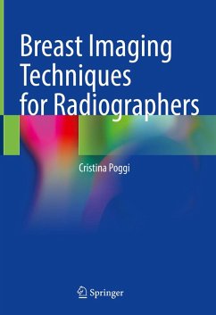
It is structured in 7 parts: 1) Basic theory on anatomy, breast cancer, treatment, and organisational features of breast screening program; 2) Technical quality, on the mammographic unit and image; 3) Clinical quality, mammography positioning, and quality assessment; 4) on the report, digital breast tomosynthesis (DBT), stereotaxis, artifacts, surgical specimen management, contrast enhancement spectral mammography (CESM), molecular breast imaging (MBI), radioprotection in the Senology department and breast magnetic resonance imaging (BMRI); 5) Ergonomics in the Senology department, radiographer academic training and communication skills; 6) advanced information in evaluating and producing high-quality mammograms, and about the Poggi method; 7) 2 annexes on clinical and technical quality in mammography and 1 about collecting historical data in Breast MRI.
Great importance is attached to the image produced, characterized by a very high level of diagnostic information, to enable the reader to find the lesion as early as possible. The question of image production is addressed, as is the thorough and appropriate assessment of its quality.
This book contains up-to-date information on breast imaging and the breast cancer patient's surveillance pathway. It will therefore be of interest to radiographers, technologists, radiologists, breast nurses and radiography students at undergraduate and postgraduate levels.
Dieser Download kann aus rechtlichen Gründen nur mit Rechnungsadresse in A, B, BG, CY, CZ, D, DK, EW, E, FIN, F, GR, HR, H, IRL, I, LT, L, LR, M, NL, PL, P, R, S, SLO, SK ausgeliefert werden.

