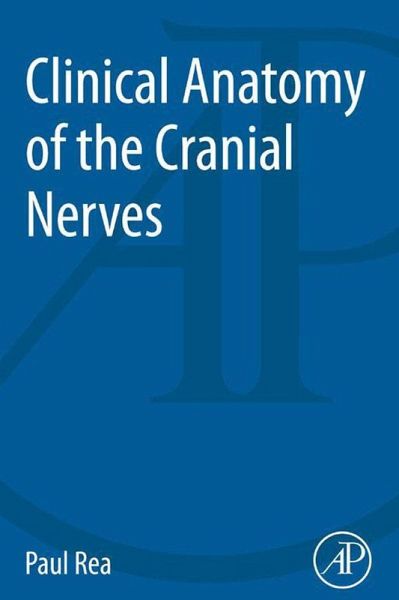
Clinical Anatomy of the Cranial Nerves (eBook, ePUB)
Versandkostenfrei!
Sofort per Download lieferbar
39,95 €
inkl. MwSt.
Weitere Ausgaben:

PAYBACK Punkte
20 °P sammeln!
Clinical Anatomy of the Cranial Nerves combines anatomical knowledge, pathology, clinical examination, and explanation of clinical findings, drawing together material typically scattered throughout anatomical textbooks. All of the pertinent anatomical topics are conveniently organized to instruct on anatomy, but also on how to examine the functioning of this anatomy in the patient. Providing a clear and succinct presentation of the underlying anatomy, with directly related applications of the anatomy to clinical examination, the book also provides unique images of anatomical structures of plas...
Clinical Anatomy of the Cranial Nerves combines anatomical knowledge, pathology, clinical examination, and explanation of clinical findings, drawing together material typically scattered throughout anatomical textbooks. All of the pertinent anatomical topics are conveniently organized to instruct on anatomy, but also on how to examine the functioning of this anatomy in the patient. Providing a clear and succinct presentation of the underlying anatomy, with directly related applications of the anatomy to clinical examination, the book also provides unique images of anatomical structures of plastinated cadaveric dissections. These images are the only ones that exist in this form, and have been professionally produced in the Laboratory of Human Anatomy, University of Glasgow under the auspices of the author. These specimens offer a novel way of visualizing the cranial nerves and related important anatomical structures. - Anatomy of cranial nerves described in text format with accompanying high-resolution images of professional, high-quality prosected cadaveric material, demonstrating exactly what the structures (and related ones) look like - Succinct yet comprehensive format with quick and easy access to facts in clearly laid out key regions, common throughout the different cranial nerves - Includes clinical examination and related pathologies, featuring diagnostic summaries of potential clinical presentations and clinically relevant questions on the anatomy of these nerves
Dieser Download kann aus rechtlichen Gründen nur mit Rechnungsadresse in A, B, BG, CY, CZ, D, DK, EW, E, FIN, F, GR, HR, H, IRL, I, LT, L, LR, M, NL, PL, P, R, S, SLO, SK ausgeliefert werden.


