-21%11
27,95 €
35,30 €**
27,95 €
inkl. MwSt.
**Preis der gedruckten Ausgabe (Broschiertes Buch)
Sofort per Download lieferbar
payback
14 °P sammeln
-21%11
27,95 €
35,30 €**
27,95 €
inkl. MwSt.
**Preis der gedruckten Ausgabe (Broschiertes Buch)
Sofort per Download lieferbar

Alle Infos zum eBook verschenken
payback
14 °P sammeln
Als Download kaufen
35,30 €****
-21%11
27,95 €
inkl. MwSt.
**Preis der gedruckten Ausgabe (Broschiertes Buch)
Sofort per Download lieferbar
payback
14 °P sammeln
Jetzt verschenken
35,30 €****
-21%11
27,95 €
inkl. MwSt.
**Preis der gedruckten Ausgabe (Broschiertes Buch)
Sofort per Download lieferbar

Alle Infos zum eBook verschenken
payback
14 °P sammeln
  • Format: PDF

Content-based image retrieval (CBIR) is the process of retrieval of images from a database that are similar to a query image, using measures derived from the images themselves, rather than relying on accompanying text or annotation. To achieve CBIR, the contents of the images need to be characterized by quantitative features; the features of the query image are compared with the features of each image in the database and images having high similarity with respect to the query image are retrieved and displayed. CBIR of medical images is a useful tool and could provide radiologists with…mehr

Produktbeschreibung
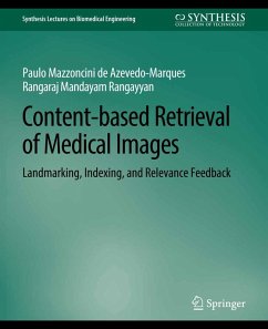
Content-based image retrieval (CBIR) is the process of retrieval of images from a database that are similar to a query image, using measures derived from the images themselves, rather than relying on accompanying text or annotation. To achieve CBIR, the contents of the images need to be characterized by quantitative features; the features of the query image are compared with the features of each image in the database and images having high similarity with respect to the query image are retrieved and displayed. CBIR of medical images is a useful tool and could provide radiologists with assistance in the form of a display of relevant past cases. One of the challenging aspects of CBIR is to extract features from the images to represent their visual, diagnostic, or application-specific information content. In this book, methods are presented for preprocessing, segmentation, landmarking, feature extraction, and indexing of mammograms for CBIR. The preprocessing steps include anisotropic diffusion and the Wiener filter to remove noise and perform image enhancement. Techniques are described for segmentation of the breast and fibroglandular disk, including maximum entropy, a moment-preserving method, and Otsu's method. Image processing techniques are described for automatic detection of the nipple and the edge of the pectoral muscle via analysis in the Radon domain. By using the nipple and the pectoral muscle as landmarks, mammograms are divided into their internal, external, upper, and lower parts for further analysis. Methods are presented for feature extraction using texture analysis, shape analysis, granulometric analysis, moments, and statistical measures. The CBIR system presented provides options for retrieval using the Kohonen self-organizing map and the k-nearest-neighbor method. Methods are described for inclusion of expert knowledge to reduce the semantic gap in CBIR, including the query point movement method for relevance feedback (RFb). Analysis of performance is described in terms of precision, recall, and relevance-weighted precision of retrieval. Results of application to a clinical database of mammograms are presented, including the input of expert radiologists into the CBIR and RFb processes. Models are presented for integration of CBIR and computer-aided diagnosis (CAD) with a picture archival and communication system (PACS) for efficient workflow in a hospital. Table of Contents: Introduction to Content-based Image Retrieval / Mammography and CAD of Breast Cancer / Segmentation and Landmarking of Mammograms / Feature Extraction and Indexing of Mammograms / Content-based Retrieval of Mammograms / Integration of CBIR and CAD into Radiological Workflow

Dieser Download kann aus rechtlichen Gründen nur mit Rechnungsadresse in A, B, BG, CY, CZ, D, DK, EW, E, FIN, F, GR, HR, H, IRL, I, LT, L, LR, M, NL, PL, P, R, S, SLO, SK ausgeliefert werden.

Autorenporträt
Paulo Mazzoncini de Azevedo-Marques is a full-time Associate Professor of Medical Physics and Biomedical Informatics with the Internal Medicine Department, University of São Paulo (USP), School of Medicine, in Ribeirão Preto, SP, Brazil. He received his B.Sc. and M.Sc. degrees in Electrical Engineering in 1986 and 1990, respectively, and his Ph.D. in Applied Physics in 1994, from USP. He has previously worked on medical imaging quality control; since 1996, his research has focused on medical image processing. He held a research associate position at the University of Chicago in 2001, where he worked on medical image processing for computer-aided diagnosis (CAD) and content-based image retrieval (CBIR), under the supervision of Professor Kunio Doi. His main subject interest areas are CAD, CBIR, and picture archival and communication systems (PACS). Rangaraj Mandayam Rangayyan is a Professor with the Department of Electrical and Computer Engineering, and an Adjunct Professor of Surgery and Radiology, at the University of Calgary, Calgary, Alberta, Canada. He received the Bachelor of Engineering degree in Electronics and Communication in 1976 from the University of Mysore at the People's Education Society College of Engineering, Mandya, Karnataka, India, and the Ph.D. degree in Electrical Engineering from the Indian Institute of Science, Bangalore, Karnataka, India, in 1980. His research interests are in the areas of digital signal and image processing, biomedical signal analysis, biomedical image analysis, and computer-aided diagnosis. He has published more than 150 papers in journals and 250 papers in proceedings of conferences. His research productivity was recognized with the 1997 and 2001 Research Excellence Awards of the Department of Electrical and Computer Engineering, the 1997 Research Award of the Faculty of Engineering, and by appointment as a "University Professor" in 2003, at the University of Calgary. He is the author of two textbooks: Biomedical Signal Analysis (IEEE/Wiley, 2002) and Biomedical Image Analysis (CRC, 2005).He has coauthored and coedited several other books, including one on Color Image Processing with Biomedical Applications (SPIE, 2011). He was recognized by the IEEE with the award of the Third Millennium Medal in 2000, and was elected as a Fellow of the IEEE in 2001, Fellow of the Engineering Institute of Canada in 2002, Fellow of the American Institute for Medical and Biological Engineering in 2003, Fellow of SPIE: the International Society for Optical Engineering in 2003, Fellow of the Society for Imaging Infor[1]matics in Medicine in 2007, Fellow of the Canadian Medical and Biological Engineering Society in 2007, and Fellow of the Canadian Academy of Engineering in 2009. He has been awarded the Killam Resident Fellowship thrice (1998, 2002, and 2007) in support of his book-writing projects.