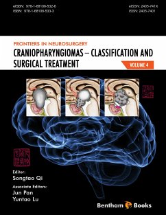
Craniopharyngiomas are a type of brain tumour in the suprasellar region with benign histological and cellular features. Clinical manifestations of craniopharyngiomas include decreased vision, hypopituitarism, hypothalamus dysfunction, fluid and electrolyte imbalance, glycometabolism, lipid metabolism and obesity. Craniopharyngiomas can show some characteristics of malignant tumours, such as invasion to the surrounding tissues, repetitious recurrence, and rapid growth. These characteristics cause considerable difficulty in patient treatment for neurosurgeons all over the world. This volume presents detailed information about craniopharyngioma anatomy, classification and treatment.
Key features:
- Detailed references based on the clinical data of more than 400 craniopharyngioma cases, and cadaveric anatomical findings of the relationship between tumours and surrounding structures.
- A new proposed topographic classification of craniopharyngiomas explained with corresponding surgical techniques for treatment.
- Analytical presentation with references to medical literature.
- Information about quality of life (QOL) and endocrinology evaluation relevant to craniopharyngioma treatment
- 30 typical clinical cases with more than 500 clear anatomical pictures, radiological and intra-surgical images, and delicate schematic and ideographic diagrams.
This reference is an essential information resource on craniopharyngiomas for neurosurgeons (adult and pediatric), neuro-endocrinologists, neuro-radiologists, ophthalmologists and medical residents.
Dieser Download kann aus rechtlichen Gründen nur mit Rechnungsadresse in A, B, BG, CY, CZ, D, DK, EW, E, FIN, F, GR, H, IRL, I, LT, L, LR, M, NL, PL, P, R, S, SLO, SK ausgeliefert werden.