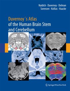
This atlas instills a solid knowledge of anatomy by correlating thin-section brain anatomy with corresponding clinical magnetic resonance images in axial, coronal, and sagittal planes. The authors correlate advanced neuromelanin imaging, susceptibility-weighted imaging, and diffusion tensor tractography with clinical 3 and 4 T MRI. Each brain stem region is then analyzed with 9.4 T MRI to show the anatomy of the medulla, pons, midbrain, and portions of the diencephalonin with an in-plane resolution comparable to myelin- and Nissl-stained light microscopy. The book's carefully organized diagrams and images teach with a minimum of text.
Dieser Download kann aus rechtlichen Gründen nur mit Rechnungsadresse in A, B, BG, CY, CZ, D, DK, EW, E, FIN, F, GR, HR, H, IRL, I, LT, L, LR, M, NL, PL, P, R, S, SLO, SK ausgeliefert werden.

