-23%11
40,95 €
53,49 €**
40,95 €
inkl. MwSt.
**Preis der gedruckten Ausgabe (Broschiertes Buch)
Sofort per Download lieferbar
payback
20 °P sammeln
-23%11
40,95 €
53,49 €**
40,95 €
inkl. MwSt.
**Preis der gedruckten Ausgabe (Broschiertes Buch)
Sofort per Download lieferbar

Alle Infos zum eBook verschenken
payback
20 °P sammeln
Als Download kaufen
53,49 €****
-23%11
40,95 €
inkl. MwSt.
**Preis der gedruckten Ausgabe (Broschiertes Buch)
Sofort per Download lieferbar
payback
20 °P sammeln
Jetzt verschenken
53,49 €****
-23%11
40,95 €
inkl. MwSt.
**Preis der gedruckten Ausgabe (Broschiertes Buch)
Sofort per Download lieferbar

Alle Infos zum eBook verschenken
payback
20 °P sammeln
  • Format: PDF

Among medical imaging modalities, magnetic resonance imaging (MRI) stands out for its excellent soft-tissue contrast, anatomical detail, and high sensitivity for disease detection. However, as proven by the continuous and vast effort to develop new MRI techniques, limitations and open challenges remain. The primary source of contrast in MRI images are the various relaxation parameters associated with the nuclear magnetic resonance (NMR) phenomena upon which MRI is based. Although it is possible to quantify these relaxation parameters (qMRI) they are rarely used in the clinic, and radiological…mehr

Produktbeschreibung
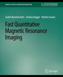
Among medical imaging modalities, magnetic resonance imaging (MRI) stands out for its excellent soft-tissue contrast, anatomical detail, and high sensitivity for disease detection. However, as proven by the continuous and vast effort to develop new MRI techniques, limitations and open challenges remain. The primary source of contrast in MRI images are the various relaxation parameters associated with the nuclear magnetic resonance (NMR) phenomena upon which MRI is based. Although it is possible to quantify these relaxation parameters (qMRI) they are rarely used in the clinic, and radiological interpretation of images is primarily based upon images that are relaxation time weighted. The clinical adoption of qMRI is mainly limited by the long acquisition times required to quantify each relaxation parameter as well as questions around their accuracy and reliability. More specifically, the main limitations of qMRI methods have been the difficulty in dealing with the high inter-parameter correlations and a high sensitivity to MRI system imperfections.

Recently, new methods for rapid qMRI have been proposed. The multi-parametric models at the heart of these techniques have the main advantage of accounting for the correlations between the parameters of interest as well as system imperfections. This holistic view on the MR signal makes it possible to regress many individual parameters at once, potentially with a higher accuracy. Novel, accurate techniques promise a fast estimation of relevant MRI quantities, including but not limited to longitudinal (T1) and transverse (T2) relaxation times. Among these emerging methods, MR Fingerprinting (MRF), synthetic MR (syMRI or MAGIC), and T1¿T2 Shuffling are making their way into the clinical world at a very fast pace. However, the main underlying assumptions and algorithms used are sometimes different from those found in the conventional MRI literature, and can be elusive at times. In this book, we take the opportunity to study and describe the main assumptions, theoretical background, and methods that are the basis of these emerging techniques.

Quantitative transient state imaging provides an incredible, transformative opportunity for MRI. There is huge potential to further extend the physics, in conjunction with the underlying physiology, toward a better theoretical description of the underlying models, their application, and evaluation to improve the assessment of disease and treatment efficacy.


Dieser Download kann aus rechtlichen Gründen nur mit Rechnungsadresse in A, B, BG, CY, CZ, D, DK, EW, E, FIN, F, GR, HR, H, IRL, I, LT, L, LR, M, NL, PL, P, R, S, SLO, SK ausgeliefert werden.

Autorenporträt
Guido Buonincontri, Ph.D., is a physicist who has been working in the field of MRI for approximately 10 years, alternating between pre-clinical and clinical MRI research. For his Ph.D. program and for a following post-doc he worked on the development and application of novel MRI techniques for cardiovascular assessments in small animals at the University of Cambridge. As a physicist working at the Department of Clinical Neuroscience first and then at the Department of Medicine, his role was to build on the latest MRI techniques to enable studies in basic science and pharmacology, with a main focus on cardiac applications. Currently, he works in Tuscany, Italy, for Fondazione Stella Maris and Fondazione Imago7, two non-profit research foundations part of a children's hospital with close ties to the University of Pisa. Guido has been a Young Investigator Fellow for INFN CSV, as well as a EU Horizon 2020 Marie-Curie Individual Fellow, focussing on fast and quantitative MRI. He is currently conducting an Italian Ministry of Health funded clinical trial assessing fast quantitative MRI in children and challenging adults and is enjoying interdisciplinary and international collaborations.
Joshua D. Kaggie, Ph.D., is a physicist and researcher at the University of Cambridge in the Department of Radiology. Born near Salt Lake City, Utah, U.S., Dr. Kaggie moved to Cambridge in the UK following his Ph.D. Dr. Kaggie did undergraduate research in gamma ray astronomy, which included the manufacture of electronics and the simulation of Cerenkov showers. Dr. Kaggie then moved into Magnetic Resonance Imaging (MRI) research for his Ph.D., where his primary focus was the development of MRI techniques for sodium imaging, which included the development of MRI transmit/receive equipment, simultaneous multinuclear imaging methods, and non-Cartesian acquisition/reconstruction. At the University of Cambridge, Dr. Kaggie has been primarily funded by GlaxoSmithKline, the Biomedical Research Council, and a European Union Horizon 2020 grant. The collaboration between all three authors began as a result of a Royal Society travel grant, when Dr. Kaggie arrived at Cambridge and as Dr. Buonincontri was leaving Cambridge for Pisa. This collaboration has proven very useful for the development of software and ideas between centers.
Martin Graves is a Consultant Clinical Scientist at Cambridge University Hospitals with over 35 years' experience in both clinical and research aspects of MRI. He received his B.Sc. (1984) and M.Sc. (1987) from the University of London and his Ph.D. (2010) from the University of Cambridge. He is a Fellow of the International Society for Magnetic Resonance in Medicine (ISMRM), the UK Institute of Physics and Engineering in Medicine (IPEM). He has been awarded Honorary Membership of the UK Royal College of Radiologists (RCR) (2016) and the IPEM Academic Gold Medal (2018). He has co-authored over 200 peer-reviewed publications as well as co-authoring a number of textbooks including MRI: From Picture to Proton (CUP 2003, 2007, and 2016), Physics MCQs for the Part 1 FRCR (CUP, 2011), and The Physics and Mathematics of MRI (Morgan & Claypool Publishers, 2016).