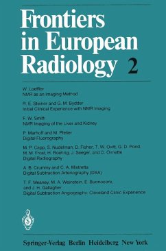
The second volume of Frontiers in European Radiology covers two very promising techniques in diagnostic radiology, namely digital radiography and nuclear mag netic resonance imaging. Leading experts in both fields from Europe and the Unit ed States were invited to give a critical overview; digital fluoroscopy is reported on mainly by American scientists since this technique has been developed primarily in the United States, while the results of nuclear magnetic resonance imaging are pre sented by British groups currently at the forefront of research in this field. The pa pers reflect the state of the art at mid-1981, when the contributors gathered for the yearly symposium on Current Topics in Diagnostic Radiology in Berne, Switzer land. Nuclear magnetic resonance imaging, also known as spin imaging or zeugmato graphy, has produced striking progress within the past few years - even within the past few months - as described in three papers of this volume. The images generally reflect the distribution of mobile protons contained within water and fats, and pro vide remarkable discrimination between different tissues. Malignant tissue might be identified with this technique, and a wide range of disorders associated with water concentration, diffusion, and flow would be amenable to study; the measurement of blood flow could be particularly interesting.
Dieser Download kann aus rechtlichen Gründen nur mit Rechnungsadresse in A, B, BG, CY, CZ, D, DK, EW, E, FIN, F, GR, HR, H, IRL, I, LT, L, LR, M, NL, PL, P, R, S, SLO, SK ausgeliefert werden.

