106,95 €
106,95 €
inkl. MwSt.
Sofort per Download lieferbar
payback
53 °P sammeln
106,95 €
106,95 €
inkl. MwSt.
Sofort per Download lieferbar

Alle Infos zum eBook verschenken
payback
53 °P sammeln
Als Download kaufen
106,95 €
inkl. MwSt.
Sofort per Download lieferbar
payback
53 °P sammeln
Jetzt verschenken
106,95 €
inkl. MwSt.
Sofort per Download lieferbar

Alle Infos zum eBook verschenken
payback
53 °P sammeln
  • Format: PDF

The period between 1950 and 1980 were the golden It also provides unique insights into how pathological years of transmission electron microscopy and pro- processes affect cell organization. duced a plethora of new information on the structure of This information is vital to current work in which cells that was coupled to and followed by biochemical the emphasis is on integrating approaches from p- and functional studies. TEM was king and each micro- teomics, molecular biology, molecular imaging and graph of a new object produced new information that physiology, and pathology to understand…mehr

  • Geräte: PC
  • ohne Kopierschutz
  • eBook Hilfe
  • Größe: 123.9MB
Produktbeschreibung
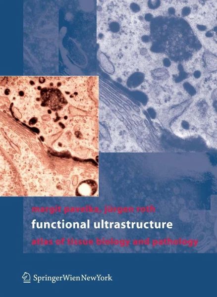
The period between 1950 and 1980 were the golden It also provides unique insights into how pathological years of transmission electron microscopy and pro- processes affect cell organization. duced a plethora of new information on the structure of This information is vital to current work in which cells that was coupled to and followed by biochemical the emphasis is on integrating approaches from p- and functional studies. TEM was king and each micro- teomics, molecular biology, molecular imaging and graph of a new object produced new information that physiology, and pathology to understand cell functions led to new insights on cell and tissue organization and and derangements in disease. In this current era, there is their functions. The quality of data represented by the a growing tendency to substitute modern light mic- images of cells and tissues had been perfected to a very scopic techniques for electron microscopy because it is high level by the great microscopists of the era including less technically demanding and is more readily available Palade, Porter, Fawcett, Sjostrand, Rhodin and many to researchers. This atlas reminds us that the infor- others. At present, the images that we see in leading tion obtained by electron microscopy is invaluable and journals for the most part do not reach the same techni- has no substitute.

Dieser Download kann aus rechtlichen Gründen nur mit Rechnungsadresse in A, B, BG, CY, CZ, D, DK, EW, E, FIN, F, GR, HR, H, IRL, I, LT, L, LR, M, NL, PL, P, R, S, SLO, SK ausgeliefert werden.

Autorenporträt
Professor Margit Pavelka, MD Studies in Medicine at the University of Vienna. Medical training at the Vienna Hospital "Rudolfstiftung" and at the Vienna General Hospital; specialization in Internal Medicine Resident at the Institute of Micromorphology and Electron Microscopy, University of Vienna; specialization in the fields of electron microscopy, cytochemistry, cytology and ultrastructural histology. Habilitation in Histology and Embryology Professor of Histology and Embryology at the Leopold Franzens University of Innsbruck and Medical University of Vienna. Head of the Center for Anatomy and Cell Biology at the Medical University of Vienna. Emerita Professor since October 2013 Professor Jürgen Roth, MD, PhD, MD hon. Studies in Medicine, Friedrich-Schiller-University, Jena Resident, Institute of Pathology, Friedrich-Schiller-University, Jena Habilitation and University Docent in Pathology, Friedrich-Schiller-University, Jena Research Associate, Department of Morphology, University of Geneva Associate Professor of Cell Biology, Biocenter, University of Basel Professor of Cell and Molecular Pathology, University of Zurich Distinguished Professor, Yonsei University Graduate School, Seoul Emeritus Professor, University of Zurich, since 2009 Editor-in-Chief of Histochemistry and Cell Biology In 2013, The Journal of Cell Biology named Jürgen Roth: Immunogold master
Rezensionen
"This is a collection of high-quality images correlated with comprehensive and detailed text to understand the ultrastructural organization of cells and several tissues. ... This book is appropriate for a broad range of audiences with the goal of providing them with information about the major role that ultrastructural analysis and novel methods such as FIB-SEM (focused ion beam-scanning electron microcopy) play in the field of cell and tissue biology and pathology." (Michele Fornaro, Doody's Book Reviews, October, 2015)