48,95 €
48,95 €
inkl. MwSt.
Sofort per Download lieferbar
payback
24 °P sammeln
48,95 €
48,95 €
inkl. MwSt.
Sofort per Download lieferbar

Alle Infos zum eBook verschenken
payback
24 °P sammeln
Als Download kaufen
48,95 €
inkl. MwSt.
Sofort per Download lieferbar
payback
24 °P sammeln
Jetzt verschenken
48,95 €
inkl. MwSt.
Sofort per Download lieferbar

Alle Infos zum eBook verschenken
payback
24 °P sammeln
  • Format: ePub

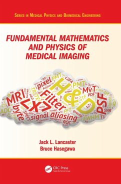
Authored by a leading educator, this book teaches the fundamental mathematics and physics concepts associated with medical imaging systems. Going beyond mere description of imaging modalities, this book delves into the mechanisms of image formation and image quality common to all imaging systems: contrast mechanisms, noise, and spatial and temporal resolution, making it an important reference for medical physicists and biomedical engineering students. This is an extensively revised new edition of The Physics of Medical X-Ray Imaging by Bruce Hasegawa (Medical Physics Publishing, 1991), and…mehr

  • Geräte: eReader
  • mit Kopierschutz
  • eBook Hilfe
  • Größe: 20.74MB
Produktbeschreibung
Authored by a leading educator, this book teaches the fundamental mathematics and physics concepts associated with medical imaging systems. Going beyond mere description of imaging modalities, this book delves into the mechanisms of image formation and image quality common to all imaging systems: contrast mechanisms, noise, and spatial and temporal resolution, making it an important reference for medical physicists and biomedical engineering students. This is an extensively revised new edition of The Physics of Medical X-Ray Imaging by Bruce Hasegawa (Medical Physics Publishing, 1991), and includes a wide range of modalities such as X-ray CT, MRI and SPECT.


Dieser Download kann aus rechtlichen Gründen nur mit Rechnungsadresse in A, B, BG, CY, CZ, D, DK, EW, E, FIN, F, GR, HR, H, IRL, I, LT, L, LR, M, NL, PL, P, R, S, SLO, SK ausgeliefert werden.

Autorenporträt
Jack Lancaster's research activities focus on theory, acquisition, and processing of 3D images of humans and animals. His research in modeling has lead to the development of new 3D standardized models of the human brain, methods to assess myelin levels during early brain development, and a new theory of interactions of pulsed E-M fields in the brain. He is a member of the Committees on Graduate Studies for the joint UTSA-UTHSCSA Biomedical Engineering Program and the UTHSCSA Radiological Sciences program, where he teaches courses in advanced imaging concepts. He is also Co-Editor of the Human Brain Mapping journal and actively involved in groups developing standards for brain mapping. He has published over 150 articles in peerreviewed journals.