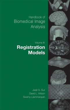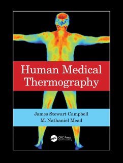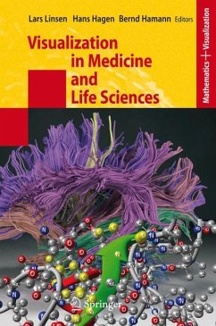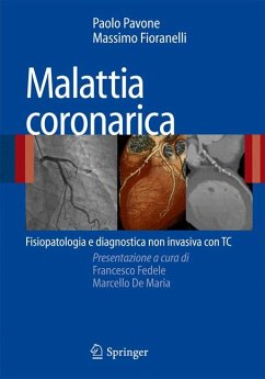
Handbook of Biomedical Image Analysis: Segmentation Models (Volume II) is dedicated to the segmentation of complex shapes from the field of imaging sciences using different mathematicaltechniques. This volume is aimed at researchers and educators in imaging sciences, radiological imaging, clinical and diagnostic imaging, physicists covering different medical imaging modalities, as well as researchers in biomedical engineering, applied mathematics, algorithmic development, computer vision, signal processing, computer graphics and multimedia in general, both in academia and industry.
Key Features:
--- Model-based brain tissue classification
--- Supervised texture classification for intravascular tissue characterization
--- Medical image segmentation: methods and applications in functional imaging
--- Automatic segmentation of pancreatic tumors in CT
--- Computerized analysis and vasodilation parameterization in flow-mediated dilation tests from ultrasonic image sequences
--- Adaptive approaches to optical segmentation in brain MRI Images
--- Automatic analysis of color fundus photographs and its application to the diagnosis of diabetic retinopathy
--- Segmentation issues of carotid plaque analysis in MRI
--- Accurate lumen identification, detection, and quantification in MR plaque volumes
--- Hessian-based multiscale enhancement, description, and quantification of second-order 3D local structures from medical volume data
--- A knowledge-based scheme for digital mammography
--- Simultaneous fuzzy segmentation
--- CAD of mammographic calicification: impact of segmentation
About the Editors:
Jasjit Suri, Ph.D. has spent over 20 years in the field of computer and electrical engineering, and more than a decade in imaging sciences. Dr. Suri has a masters degree in computer sciences from the University of Illinois and a doctorate in Electrical Engineering from the University of Washington, Seattle. Dr. Suri has published over 125 technical publications in medical imaging, as well as being a senior member of IEEE and a member of the engineering honor societies Eta-Kappa-Nu and Tau-Beta-Phi, and a recipient of the President's Gold Medal in 1980. He is also a fellow of American Institute of Medical and Biological Engineering.
David Wilson, Ph D. is a Professor of Biomedical Engineering and Radiology at Case Western Reserve University, having gained his doctorate from Rice University. He has over 60 refereed journal publications and is co-owner of several patents. Professor Wilson has actively developed biomedical imaging at CWRU. He has led a faculty recruitment effort, and he has served as PI or has been an active leader on multiple research and equipment developmental awards given to CWRU, including an NIH planning grant award for an in vivo Cellular and Molecular Imaging Center and an Ohio Wright Center of Innovation award. Swamy Laxminarayan, Dsc championed the field of Biomedical Engineering for over 30 years, having held a variety of senior positions within the industry. He is an internationally recognized scientist, engineer, and educator and has been published in over 200 technical publications in biomedical information technology, computation biology, signal and image processing, biotechnology, and physiological system modeling. Prof. Laxminarayan is a fellow of AIMBE and a recipient of IEEE 3rd Millennium Medal.
Dieser Download kann aus rechtlichen Gründen nur mit Rechnungsadresse in A, B, BG, CY, CZ, D, DK, EW, E, FIN, F, GR, HR, H, IRL, I, LT, L, LR, M, NL, PL, P, R, S, SLO, SK ausgeliefert werden.