109,95 €
109,95 €
inkl. MwSt.
Sofort per Download lieferbar
payback
55 °P sammeln
109,95 €
109,95 €
inkl. MwSt.
Sofort per Download lieferbar

Alle Infos zum eBook verschenken
payback
55 °P sammeln
Als Download kaufen
109,95 €
inkl. MwSt.
Sofort per Download lieferbar
payback
55 °P sammeln
Jetzt verschenken
109,95 €
inkl. MwSt.
Sofort per Download lieferbar

Alle Infos zum eBook verschenken
payback
55 °P sammeln
  • Format: ePub

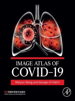
Image Atlas of COVID-19 covers this novel and emerging respiratory infectious disease. Although the gold standard for clinical diagnosis of COVID-19 remains the nucleic acid test for SARS-CoV-2, chest CT scans have been widely used in the diagnosis and treatment of COVID-19 as simple and fast diagnostic methods. The latter has proven crucial when faced with a large volume of symptomatic and asymptomatic patients. The imaging manifestations of COVID-19 have some specific characteristics, hence a better understanding of imaging findings is of great value for detecting lesions, evaluating lesion…mehr

Produktbeschreibung
Image Atlas of COVID-19 covers this novel and emerging respiratory infectious disease. Although the gold standard for clinical diagnosis of COVID-19 remains the nucleic acid test for SARS-CoV-2, chest CT scans have been widely used in the diagnosis and treatment of COVID-19 as simple and fast diagnostic methods. The latter has proven crucial when faced with a large volume of symptomatic and asymptomatic patients. The imaging manifestations of COVID-19 have some specific characteristics, hence a better understanding of imaging findings is of great value for detecting lesions, evaluating lesion severity, diagnosis and differential diagnosis, and assessment of therapeutic options and prognosis.
  • Contains 61 confirmed cases of COVID-19, including a collection of over 400 DR, CT, PET/CT and PET/MRI scans in Henan Provincial People's Hospital (HPPH)
  • Organized into four chapters by clinical subtypes of COVID-19
  • Details a large number of clinical images, with illustrations that are intended to demonstrate imaging findings of COVID-19 to serve as a reference manual for medical staff engaged in imaging and clinical work
  • Ideal as a training material for academic education

Dieser Download kann aus rechtlichen Gründen nur mit Rechnungsadresse in A, B, BG, CY, CZ, D, DK, EW, E, FIN, F, GR, HR, H, IRL, I, LT, L, LR, M, NL, PL, P, R, S, SLO, SK ausgeliefert werden.

Autorenporträt
Professor Wang M.D., Ph.D serves as Board of Trustee (BoT) member of the International Society for Magnetic Resonance in Medicine (ISMRM),Certified Teacher in Clinical MRI of ISMRM and European Society for Magnetic Resonance in Medicine and Biology (ESMRMB), Vice-Chair of the Committee on Women in Chinese Society of Biomedical Engineering (CSBME) and standing member of the Artificial Intelligence Committee in CSBME. Also, she is the President-Elect of the International Society for Neurovascular Disease (ISNVD) and Overseas Chinese Society for Magnetic Resonance in Medicine (OCSMRM), Vice-Chair of the Chinese Chapter of the ISMRM, and she has served on the Annual Meeting Program Committee (AMPC), the MR Value Initiative Committee, the Chapters Committee and the Ad Hoc Committee on Women in ISMRM, Secretary and Chair in 2021 of the Psychiatric MR Spectroscopy & Imaging Study Group of ISMRM. She has had two years' postdoc in A.A. Martinos Center for Biomedical Imaging, Massachusetts General Hospital, Harvard Medical School from 2006 to 2008, and twenty-five years' working experience in diagnostic MR imaging and MRI research. Her research has focused on exploring the value of multi-model imaging including CT, MRI, PET-CT, PET-MR, and MR-guided Focused Ultrasound (MRgFUS) to improve diagnosis and treatment of diseases. Dr. Wang has published over 100 papers on journals such as JAMA, Molecular Psychiatry, Radiology and given over 300 invited lectures at international and national scientific and educational meetings. She is also the principal investigator on international cooperative grants from the National Natural Science Foundation of China and the National Key R&D Program of China. Owing to her contributions to the field, she was awarded the 2018 Outstanding Contribution Award by the Overseas Chinese Society for Magnetic Resonance in Medicine (OCSMRM) and the Gold Medal of the Chinese Society of Radiology.Professor El Fakhri is the Nathaniel & Diana Alpert Professor of Radiology at Harvard Medical School (HMS) and the founding Director of the Gordon Center for Medical Imaging at Massachusetts General Hospital and HMS with over 150 members. He is also co-Director of the Division of Nuclear Medicine and Molecular Imaging and Associate Chair for Medical Sciences of the Department of Radiology at MGH. Dr. El Fakhri is an internationally recognized expert in quantitative molecular imaging (SPECT, PET-CT, and PET-MR) for in vivo assessment of patho-physiology in brain, cardiac and oncologic diseases. Current areas of research include high resolution PET & MR imaging in a range of diseases including neurodegenerative disease and traumatic brain injury (amyloid and neurofibrillary tangles), cardiac disease (mitochondrial membrane potential), as well as guiding radiotherapy planning (PET/MRS). He has authored or co-authored over 300 papers and mentored over 100 students, post-docs and faculty and has delivered over 200 invited Talks, Keynote and Plenary Lectures. Dr. El Fakhri received many awards and honors, including the Mark Tetalman Award from the Society of Nuclear Medicine, the Dana Foundation Brain and Immuno-Imaging Award, the Howard Hughes Medical Institutes Training Innovation Award and the Edward J. Hoffman Award from the Society of Nuclear Medicine and Molecular Imaging. He was elected Fellow to the SNMMI, AAPM, AIMBE, and IEEE for "contributions to biological imaging.