73,95 €
73,95 €
inkl. MwSt.
Sofort per Download lieferbar
payback
37 °P sammeln
73,95 €
73,95 €
inkl. MwSt.
Sofort per Download lieferbar

Alle Infos zum eBook verschenken
payback
37 °P sammeln
Als Download kaufen
73,95 €
inkl. MwSt.
Sofort per Download lieferbar
payback
37 °P sammeln
Jetzt verschenken
73,95 €
inkl. MwSt.
Sofort per Download lieferbar

Alle Infos zum eBook verschenken
payback
37 °P sammeln
  • Format: PDF

Patients with gliomas frequently undergo combination therapy that can result in complex and potentially confusing appearances on follow-up imaging. In this context, differentiation between tumor recurrence and radiation necrosis, for example, can be very difficult.
This atlas is a detailed guide to the imaging appearances of gliomas following treatment with neurosurgery, radiation therapy, and chemotherapy. Normal and pathological findings are displayed in detailed MR images that illustrate the potential modifications due to treatment. Particular emphasis is placed on characteristic…mehr

  • Geräte: PC
  • ohne Kopierschutz
  • eBook Hilfe
  • Größe: 12.87MB
Produktbeschreibung
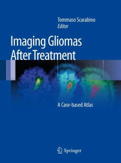
Patients with gliomas frequently undergo combination therapy that can result in complex and potentially confusing appearances on follow-up imaging. In this context, differentiation between tumor recurrence and radiation necrosis, for example, can be very difficult.

This atlas is a detailed guide to the imaging appearances of gliomas following treatment with neurosurgery, radiation therapy, and chemotherapy. Normal and pathological findings are displayed in detailed MR images that illustrate the potential modifications due to treatment. Particular emphasis is placed on characteristic appearances on the newer functional MR imaging techniques, including MR spectroscopy, diffusion-weighted imaging, and perfusion imaging. These techniques are revolutionizing neuroradiology by going beyond the demonstration of macroscopic alterations to the depiction of preceding metabolic changes at the cellular and subcellular level. They thereby improve the accuracy, sensitivity, and specificity of MR imaging and allow earlier and more specific diagnosis. A key section comprising some 40 clinical cases and more than 500 illustrations offers an invaluable clinical and research tool not only for neuroradiologists but also for neurosurgeons, radiotherapists, and medical oncologists.


Dieser Download kann aus rechtlichen Gründen nur mit Rechnungsadresse in A, B, BG, CY, CZ, D, DK, EW, E, FIN, F, GR, HR, H, IRL, I, LT, L, LR, M, NL, PL, P, R, S, SLO, SK ausgeliefert werden.