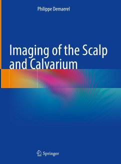
This richly illustrated book provides a comprehensive account of the imaging of scalp and calvarial lesions. It discusses essential facts such as the anatomy and pathology of the scalp and calvarium, imaging findings in CT and MRI, differential diagnosis, and selected references. The author presents the key information on the left and illustrations on the right side of the book. While the book shows the most common radiological examples, it also includes less typical cases.
The uniform design and easy-to-use structure make the book a valuable reference guide for (neuro)radiology, neurosurgery, and dermatology specialists.
Dieser Download kann aus rechtlichen Gründen nur mit Rechnungsadresse in A, B, BG, CY, CZ, D, DK, EW, E, FIN, F, GR, HR, H, IRL, I, LT, L, LR, M, NL, PL, P, R, S, SLO, SK ausgeliefert werden.


