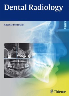
Imaging Techniques in Dental Radiology (eBook, PDF)
Acquisition, Anatomic Analysis and Interpretation of Radiographic Images
Versandkostenfrei!
Sofort per Download lieferbar
72,95 €
inkl. MwSt.
Weitere Ausgaben:

PAYBACK Punkte
36 °P sammeln!
This book is an up-to-date guide to the performance and interpretation of imaging studies in dental radiology. After opening discussion of the choice of X-ray equipment and materials, intraoral radiography, panoramic radiography, cephalometric radiology, and cone-beam computed tomography are discussed in turn. With the aid of many illustrated examples, patient preparation and positioning are thoroughly described for each modality. Common technical errors and artifacts are identified and the means of avoiding them, explained. The aim is to equip the reader with all the information required in o...
This book is an up-to-date guide to the performance and interpretation of imaging studies in dental radiology. After opening discussion of the choice of X-ray equipment and materials, intraoral radiography, panoramic radiography, cephalometric radiology, and cone-beam computed tomography are discussed in turn. With the aid of many illustrated examples, patient preparation and positioning are thoroughly described for each modality. Common technical errors and artifacts are identified and the means of avoiding them, explained. The aim is to equip the reader with all the information required in order to perform imaging effectively and safely. The normal radiographic anatomy and landmarks are then discussed, prior to thorough coverage of frequent dentomaxillofacial lesions. Accompanying images display the characteristic features of each lesion. Further topics to be addressed are safety precautions for patients and staff. The book will be an ideal aid for all dental practitioners and will also be of value for dental students.
Dieser Download kann aus rechtlichen Gründen nur mit Rechnungsadresse in A, B, BG, CY, CZ, D, DK, EW, E, FIN, F, GR, HR, H, IRL, I, LT, L, LR, M, NL, PL, P, R, S, SLO, SK ausgeliefert werden.


