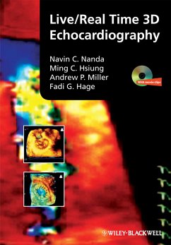
This comprehensive, state-of-the-art review of both live/real time 3D transthoracic and transesophageal echocardiography illustrates both normal and pathologic cardiovascular findings. With more than 800 images that detail the technique of performing these studies and demonstrate various cardiovascular pathologies, as well as a DVD containing more than 350 moving images, it is a valuable compendium for both novice and experienced practitioners. The book opens with chapters on the history of 3D echocardiography and basic and technical aspects of live/real time 3D transthoracic and transesophageal echocardiograpy, then considers: * normal anatomy, examination protocols, and the technique for performing live/real time 3D transthoracic echocardiography * abnormalities affecting the mitral, aortic, tricuspid, and pulmonary valves and the aorta * prosthetic heart valves * 3D echocardiographic assessment of left and right ventricular function, ischemic heart disease, and cardiomyopathies * congenital cardiac lesions * tumors and other mass lesions * pericardial disorders * live/real time 3D transesophageal echocardiography It concludes with coverage of some of the most recent advances in 3D technology, real time full-volume imaging, and 3D wall tracking, including 3D assessment of strain, strain rate, twist, and torsion. Vividly demonstrating the superiority of 3D echocardiography over conventional 2D imaging in several clinical situations, this carefully produced volume shows how to use the most recent technology for better assessment of cardiovascular disease.
Dieser Download kann aus rechtlichen Gründen nur mit Rechnungsadresse in A, B, BG, CY, CZ, D, DK, EW, E, FIN, F, GR, HR, H, IRL, I, LT, L, LR, M, NL, PL, P, R, S, SLO, SK ausgeliefert werden.

