Medical Devices (eBook, PDF)
Surgical and Image-Guided Technologies


Alle Infos zum eBook verschenken

Medical Devices (eBook, PDF)
Surgical and Image-Guided Technologies
- Format: PDF
- Merkliste
- Auf die Merkliste
- Bewerten Bewerten
- Teilen
- Produkt teilen
- Produkterinnerung
- Produkterinnerung

Hier können Sie sich einloggen

Bitte loggen Sie sich zunächst in Ihr Kundenkonto ein oder registrieren Sie sich bei bücher.de, um das eBook-Abo tolino select nutzen zu können.
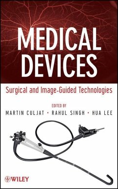
Addressing the exploding interest in bioengineering for healthcare applications, this book provides readers with detailed yet easy-to-understand guidance on biomedical device engineering. Written by prominent physicians and engineers, Medical Devices: Surgical and Image-Guided Technologies is organized into stand-alone chapters covering devices and systems in diagnostic, surgical, and implant procedures. Assuming only basic background in math and science, the authors clearly explain the fundamentals for different systems along with such topics as engineering considerations, therapeutic…mehr
- Geräte: PC
- mit Kopierschutz
- eBook Hilfe
- Größe: 7.59MB
![Medical Devices (eBook, ePUB) Medical Devices (eBook, ePUB)]() Martin CuljatMedical Devices (eBook, ePUB)136,99 €
Martin CuljatMedical Devices (eBook, ePUB)136,99 €![Medical Instrumentation (eBook, PDF) Medical Instrumentation (eBook, PDF)]() Medical Instrumentation (eBook, PDF)111,99 €
Medical Instrumentation (eBook, PDF)111,99 €![Medical Device Design for Six Sigma (eBook, PDF) Medical Device Design for Six Sigma (eBook, PDF)]() Basem El-HaikMedical Device Design for Six Sigma (eBook, PDF)167,99 €
Basem El-HaikMedical Device Design for Six Sigma (eBook, PDF)167,99 €![Imaging Systems for Medical Diagnostics (eBook, PDF) Imaging Systems for Medical Diagnostics (eBook, PDF)]() Imaging Systems for Medical Diagnostics (eBook, PDF)106,99 €
Imaging Systems for Medical Diagnostics (eBook, PDF)106,99 €![Chemical Sensors and Biosensors (eBook, PDF) Chemical Sensors and Biosensors (eBook, PDF)]() Chemical Sensors and Biosensors (eBook, PDF)160,99 €
Chemical Sensors and Biosensors (eBook, PDF)160,99 €![Tactile Sensing and Displays (eBook, PDF) Tactile Sensing and Displays (eBook, PDF)]() Javad DargahiTactile Sensing and Displays (eBook, PDF)98,99 €
Javad DargahiTactile Sensing and Displays (eBook, PDF)98,99 €![Biosensing Using Nanomaterials (eBook, PDF) Biosensing Using Nanomaterials (eBook, PDF)]() Biosensing Using Nanomaterials (eBook, PDF)171,99 €
Biosensing Using Nanomaterials (eBook, PDF)171,99 €-
-
-
Dieser Download kann aus rechtlichen Gründen nur mit Rechnungsadresse in A, B, BG, CY, CZ, D, DK, EW, E, FIN, F, GR, HR, H, IRL, I, LT, L, LR, M, NL, PL, P, R, S, SLO, SK ausgeliefert werden.
- Produktdetails
- Verlag: John Wiley & Sons
- Seitenzahl: 456
- Erscheinungstermin: 18. Oktober 2012
- Englisch
- ISBN-13: 9781118452790
- Artikelnr.: 37343428
- Verlag: John Wiley & Sons
- Seitenzahl: 456
- Erscheinungstermin: 18. Oktober 2012
- Englisch
- ISBN-13: 9781118452790
- Artikelnr.: 37343428
- Herstellerkennzeichnung Die Herstellerinformationen sind derzeit nicht verfügbar.
CONTRIBUTORS xix
PART I INTRODUCTION TO MEDICAL DEVICES 1
1. Introduction 3
Martin Culjat
1.1 History of Medical Devices 3
1.2 Medical Device Terminology 6
1.3 Purpose of the Book 10
2. Design of Medical Devices 11
Gregory Nighswonger
2.1 Introduction 11
2.2 The Medical Device Design Environment 11
2.2.1 US Regulation 12
2.2.2 Differences in European Regulation 13
2.2.3 Standards 14
2.3 Basic Design Phases 15
2.3.1 Feasibility 15
2.3.2 Planning and Organization-Assembling the Design Team 16
2.3.3 When to Involve Regulatory Affairs 17
2.3.4 Conceptualizing and Review 17
2.3.5 Testing and Refinement 20
2.3.6 Proving the Concept 20
2.3.7 Pilot Testing and Release to Manufacturing 22
2.4 Postmarket Activities 25
2.5 Final Note 25
PART II MINIMALLY INVASIVE DEVICES AND TECHNIQUES 27
3. Instrumentation for Laparoscopic Surgery 29
Camellia Racu-Keefer, Scott Um, Martin Culjat, and Erik Dutson
3.1 Introduction 29
3.2 Basic Principles 31
3.3 Laparoscopic Instrumentation 34
3.3.1 Trocars 34
3.3.2 Standard Laparoscopic Instruments 37
3.3.3 Additional Laparoscopic Instruments 42
3.3.4 Specimen Retrieval Bags 44
3.3.5 Disposable Instruments 44
3.4 Innovative Applications 45
3.5 Summary and Future Applications 46
4. Surgical Instruments in Ophthalmology 49
Allen Y. Hu, Robert M. Beardsley, and Jean-Pierre Hubschman
4.1 Introduction 49
4.2 Cataract Surgery 51
4.2.1 Basic Technique 51
4.2.2 Principles of Phacoemulsification 52
4.2.3 Phacoemulsification Instruments 54
4.2.4 Phacoemulsification Systems 55
4.2.5 Future Directions 56
4.3 Vitreoretinal Surgery 56
4.3.1 Basic Techniques 56
4.3.2 Principles of Vitrectomy 57
4.3.3 Vitrectomy Instruments 58
4.3.4 Vitrectomy Systems 60
4.3.5 Future Directions 60
4.4 Other Ophthalmic Surgical Procedures 61
4.5 Conclusion 62
5. Surgical Robotics 63
Jacob Rosen
5.1 Introduction 63
5.2 Background and Leading Concepts 63
5.2.1 Human-Machine Interfaces: System Approach 65
5.2.2 Tissue Biomechanics 70
5.2.3 Teleoperation 72
5.2.4 Image-Guided Surgery 78
5.2.5 Objective Assessment of Skill 79
5.3 Commercial Systems 80
5.3.1 ROBODOC® (Curexo Technology Corporation) 80
5.3.2 daVinci (Intuitive Surgical) 83
5.3.3 Sensei® X (Hansen Medical) 84
5.3.4 RIO® MAKOplasty (MAKO Surgical Corporation) 86
5.3.5 CyberKnife (Accuray) 89
5.3.6 Renaissance(TM) (Mazor Robotics) 91
5.3.7 ARTAS® System (Restoration Robotics, Inc.) 92
5.4 Trends and Future Directions 93
6. Catheters in Vascular Therapy 99
Axel Boese
6.1 Introduction 99
6.2 Historic Overview 100
6.3 Catheter Interventions 102
6.4 Catheter and Guide Wire Shapes and Configurations 105
6.4.1 Catheters 105
6.4.2 Guide Wires 113
6.5 Conclusion 116
PART III ENERGY DELIVERY DEVICES AND SYSTEMS 119
7. Energy-Based Hemostatic Surgical Devices 121
Amit P. Mulgaonkar, Warren Grundfest, and Rahul Singh
7.1 Introduction 121
7.2 History of Energy-Based Hemostasis 122
7.3 Energy-Based Surgical Methods and Their Effects on Tissues 125
7.3.1 Disambiguation 126
7.3.2 Thermal Effects on Tissues 127
7.4 Electrosurgery 128
7.4.1 Electrosurgical Theory 128
7.4.2 Cutting and Coagulation Techniques 130
7.4.3 Equipment 131
7.4.4 Considerations and Complications 133
7.5 Future Of Electrosurgery 134
7.6 Conclusion 135
8. Tissue Ablation Systems 137
Michael Douek, Justin McWilliams, and David Lu
8.1 Introduction 137
8.2 Evolving Paradigms in Cancer Therapy 138
8.3 Basic Ablation Categories and Nomenclature 140
8.4 Hyperthermic Ablation 140
8.5 Fundamentals of In Vivo Energy Deposition 141
8.6 Hyperthermic Ablation: Optimizing Tissue Ablation 143
8.7 Radiofrequency Ablation 144
8.8 RFA: Basic Principles 145
8.9 RFA: In Vivo Energy Deposition 145
8.10 Optimizing RFA 147
8.11 Other Hyperthermic Ablation Techniques 149
8.11.1 Microwave Ablation (MWA) 149
8.11.2 MWA: Basic Principles 149
8.11.3 MWA: In Vivo Energy Deposition 151
8.11.4 Optimizing MWA 152
8.12 Laser Ablation 153
8.13 Hypothermic Ablation 154
8.13.1 Cryoablation: Basic Concepts 154
8.13.2 Cryoablation: In Vivo Considerations 154
8.13.3 Optimizing Cryoablation Systems 154
8.14 Chemical Ablation 157
8.15 Novel Techniques 158
8.15.1 High Intensity Focused Ultrasound (HIFU) 158
8.15.2 Irreversible Electroporation (IRE) 159
8.16 Tumor Ablation and Beyond 160
9. Lasers in Medicine 163
Zachary Taylor, Asael Papour, Oscar Stafsudd, and Warren Grundfest
9.1 Introduction 163
9.1.1 Historical Perspective 164
9.1.2 Basic Operational Concepts 165
9.1.3 First Experimental MASER (Microwave Amplification by Stimulated
Emission of Radiation) 166
9.2 Laser Fundamentals 167
9.2.1 Two-Level Systems and Population Inversion 167
9.2.2 Multiple Energy Levels 167
9.2.3 Mode of Operation 169
9.2.4 Beams and Optics 171
9.3 Laser Light Compared to Other Sources of Light 174
9.3.1 Temporal Coherence 174
9.3.2 Spectral Coherence (Line Width) 175
9.3.3 Beam Collimation 177
9.3.4 Short Pulse Duration 177
9.3.5 Summary 178
9.4 Laser-Tissue Interactions 178
9.4.1 Biostimulation 178
9.4.2 Photochemical Interactions 179
9.4.3 Photothermal Interactions 180
9.4.4 Ablation 180
9.4.5 Photodisruption 181
9.5 Lasers in Diagnostics 181
9.5.1 Optical Coherence Tomography 181
9.5.2 Fluorescence Angiography 184
9.5.3 Near Infrared Spectroscopy 185
9.6 Laser Treatments and Therapy 186
9.6.1 Overview of Current Medical Applications of Laser Technology 186
9.6.2 Retinal Photodynamic Therapy (Photochemical) 188
9.6.3 Transpupillary Thermal Therapy (TTT) (Photothermal) 188
9.6.4 Vascular Birth Marks (Photocoagulation) 190
9.6.5 Laser Assisted Corneal Refractive Surgery (Ablation) 191
9.7 Conclusions 196
PART IV IMPLANTABLE DEVICES AND SYSTEMS 197
10. Vascular and Cardiovascular Devices 199
Dan Levi, Allan Tulloch, John Ho, Colin Kealey, and David Rigberg
10.1 Introduction 199
10.2 Biocompatibility Considerations 200
10.3 Materials 202
10.3.1 316L Stainless Steel 203
10.3.2 Nitinol 203
10.3.3 Cobalt-Chromium Alloys 204
10.4 Stents 204
10.5 Closure Devices 206
10.6 Transcatheter Heart Valves 208
10.7 Inferior Vena Cava Filters 212
10.8 Future Directions-Thin Film Nitinol 214
10.9 Conclusion 216
11. Mechanical Circulatory Support Devices 219
Colin Kealey, Paymon Rahgozar, and Murray Kwon
11.1 Introduction 219
11.2 History 220
11.3 Basic Principles 221
11.3.1 Biocompatibility and Mechanical Circulatory Support Devices 221
11.3.2 Hemocompatibility: Microscopic Considerations 222
11.3.3 Hemocompatibility: Macroscopic Considerations 223
11.4 Engineering Considerations in Mechanical Circulatory Support 223
11.4.1 Overview 223
11.4.2 Pump Design 225
11.4.3 Positive Displacement Pumps 225
11.4.4 Rotary Pumps 226
11.4.5 Pulsatile Versus Nonpulsatile Flow 228
11.5 Devices 228
11.5.1 The HeartMate XVE Left Ventricular Assist System 228
11.5.2 The HeartMate II Left Ventricular Assist System 231
11.5.3 Short-Term Mechanical Circulatory Support: The Intraaortic Balloon
Pump 234
11.5.4 Pediatric Mechanical Circulatory Support: The Berlin Heart 237
11.6 The Future of MCS Devices 239
11.6.1 CorAide 239
11.6.2 HeartMate III 239
11.6.3 HeartWare 240
11.6.4 VentrAssist 240
11.7 Summary 240
12. Orthopedic Implants 241
Sophia N. Sangiorgio, Todd S. Johnson, Jon Moseley, G. Bryan Cornwall, and
Edward Ebramzadeh
12.1 Introduction 241
12.1.1 Overview 241
12.1.2 History 243
12.2 Basic Principles 244
12.2.1 Optimization for Strength and Stiffness 245
12.2.2 Maximization of Implant Fixation to Host Bone 250
12.2.3 Minimization of Degradation 251
12.2.4 Sterilization of Implants and Instrumentation 253
12.3 Implant Technologies 253
12.3.1 Total Hip Replacement 254
12.3.2 Technology in Total Knee Replacement 263
12.3.3 Technology in Spine Surgery 268
12.4 Summary 272
PART V IMAGING AND IMAGE-GUIDED TECHNIQUES 275
13. Endoscopy 277
Gregory Nighswonger
13.1 Introduction 277
13.2 Ancient Origins 278
13.3 Modern Endoscopy 280
13.3.1 Creating Cold Light 280
13.3.2 Introduction of Rod-Lens Technology 280
13.4 Principles of Modern Endoscopy 283
13.4.1 Optics 284
13.4.2 Mechanics 284
13.4.3 Electronics 284
13.4.4 Software 285
13.5 The Imaging Chain 285
13.5.1 Light Source (1) 286
13.5.2 Telescope (2) 286
13.5.3 Camera Head (3) 287
13.5.4 Camera CCU (4) 287
13.5.5 Video Cables (5) 287
13.5.6 Monitor (6) 287
13.5.7 Image Management Systems (7) 288
13.6 Endoscopes for Today 288
13.6.1 Rigid Endoscopes-Designs to Enhance Functionality 289
13.6.2 Less Traumatic Ureterorenoscopes 290
13.6.3 Advances in Flexible Endoscope Design 291
13.6.4 Broader Functionality with New Technologies 294
13.6.5 Enhancing Video Capabilities 299
13.7 Endoscopy's Future 301
14. Medical Ultrasound Devices 303
Rahul Singh and Martin Culjat
14.1 Introduction 303
14.2 Basic Principles of Ultrasound 304
14.2.1 Basic Acoustic Physics 304
14.2.2 Reflection and Refraction 307
14.2.3 Attenuation 307
14.2.4 Piezoelectricity 308
14.2.5 Ultrasound Systems 310
14.2.6 Resolution and Bandwidth 312
14.2.7 Beam Characteristics 314
14.3 Ultrasound Transducer Design 316
14.3.1 Piezoelectric Material 317
14.3.2 Backing Layers and Damping 318
14.3.3 Matching Layers 318
14.3.4 Mechanical Focusing 319
14.3.5 Electrical Matching 320
14.3.6 Sector Scanners 320
14.3.7 Array Transducers 322
14.3.8 Transducer Array Fabrication 325
14.3.9 Regulatory Considerations 327
14.4 Applications of Medical Ultrasound 329
14.4.1 Image Guidance Applications 330
14.4.2 Intravascular and Intracardiac Applications 332
14.4.3 Intraoral and Endocavity Applications 333
14.4.4 Surgical Applications 334
14.4.5 Ophthalmic Ultrasound 335
14.4.6 Doppler and Doppler Applications 336
14.4.7 Therapeutic Applications 336
14.5 The Future of Medical Ultrasound 338
15. Medical X-ray Imaging 341
Mark Roden
15.1 Introduction 341
15.2 X-ray Physics 342
15.2.1 Photon Interactions with Matter 342
15.2.2 Clinical Production of X-rays 343
15.2.3 Patient Dose Considerations 346
15.3 Two-Dimensional Image Acquisition 348
15.4 Image Acquisition Technologies and Techniques 351
15.4.1 Film 351
15.4.2 Computed Radiography 354
15.4.3 Digital Radiography 358
15.4.4 Clinical Applications of 2D X-ray Techniques 360
15.5 Basic 2D Processing Techniques 361
15.5.1 Independent Pixel Operations 362
15.5.2 Grouped Pixel Operations 363
15.5.3 Image Transformation Operations 366
15.6 Real-Time X-ray Imaging 367
15.6.1 Fluoroscopy Technology 367
15.6.2 Angiography 370
15.7 Three-Dimensional X-ray Imaging 372
15.8 Conclusion 373
16. Navigation in Neurosurgery 375
Jean-Jacques Lemaire, Eric J. Behnke, Andrew J. Frew, and Antonio A. F.
DeSalles
16.1 Basics of Neurosurgery 375
16.1.1 General Technical Issues in Neurosurgery 375
16.1.2 Instrumentation in Neurosurgery 376
16.1.3 Complications 377
16.1.4 Functional Neurosurgery 378
16.1.5 Stereotactic Neurosurgery 378
16.1.6 Neuroimaging for Neurosurgery 379
16.2 Introduction to Neuronavigation 381
16.3 Neuronavigation Systems 381
16.3.1 The Tracking System 382
16.3.2 The Display Unit 383
16.3.3 The Control Unit 385
16.4 Implementation of Neuronavigation 386
16.4.1 Surgical Planning 386
16.4.2 Patient Registration 387
16.4.3 Navigation 389
16.5 Augmented Reality and Virtual Reality 390
16.6 Summary/Future 391
REFERENCES 395
INDEX 425
