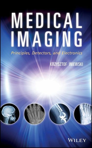
Medical Imaging (eBook, PDF)
Principles, Detectors, and Electronics
Redaktion: Iniewski, Krzysztof
Alle Infos zum eBook verschenken
Medical Imaging (eBook, PDF)
Principles, Detectors, and Electronics
Redaktion: Iniewski, Krzysztof
- Format: PDF
- Merkliste
- Auf die Merkliste
- Bewerten Bewerten
- Teilen
- Produkt teilen
- Produkterinnerung
- Produkterinnerung
Hier können Sie sich einloggen
Bitte loggen Sie sich zunächst in Ihr Kundenkonto ein oder registrieren Sie sich bei bücher.de, um das eBook-Abo tolino select nutzen zu können.
A must-read for anyone working in electronics in the healthcare sector This one-of-a-kind book addresses state-of-the-art integrated circuit design in the context of medical imaging of the human body. It explores new opportunities in ultrasound, computed tomography (CT), magnetic resonance imaging (MRI), nuclear medicine (PET, SPECT), emerging detector technologies, circuit design techniques, new materials, and innovative system approaches. Divided into four clear parts and with contributions from a panel of international experts, Medical Imaging systematically covers: * X-ray imaging and…mehr
- Geräte: PC
- mit Kopierschutz
- eBook Hilfe
- Größe: 6.87MB
Dieser Download kann aus rechtlichen Gründen nur mit Rechnungsadresse in A, B, BG, CY, CZ, D, DK, EW, E, FIN, F, GR, HR, H, IRL, I, LT, L, LR, M, NL, PL, P, R, S, SLO, SK ausgeliefert werden.
- Produktdetails
- Verlag: Wiley-Scrivener
- Seitenzahl: 328
- Erscheinungstermin: 2. März 2009
- Englisch
- ISBN-13: 9780470451809
- Artikelnr.: 37292331
- Verlag: Wiley-Scrivener
- Seitenzahl: 328
- Erscheinungstermin: 2. März 2009
- Englisch
- ISBN-13: 9780470451809
- Artikelnr.: 37292331
About The Editor xv
contributors xvii
I X-Ray Imaging and Computed Tomography 1
1 X-Ray and Computed Tomography Imaging Principles 3
Krzysztof Iniewski
1.1. Introduction to X-Ray Imaging 3
1.2. X-Ray Generation 6
1.3. X-Ray Interaction with Matter 9
1.4. X-Ray Detection 12
1.5. Electronics for X-Ray Detection 13
1.6. CT Imaging Principle 14
1.7. CT Scanners 15
1.8. Color X-Ray Imaging 17
1.9. Future of X-Ray and CT Imaging 18
References 21
2 Active Matrix Flat Panel Imagers (AMFPI) for Diagnostic Medical Imaging
Applications 23
Karim Karim
2.1. Introduction 23
2.1.1. Digital Imaging 23
2.1.2. Detection Schemes 24
2.1.3. Chapter Organization 27
2.2. Pixel Technology 27
2.2.1. Operation 27
2.2.1.1. Introduction 27
2.2.1.2. Operation 28
2.2.1.3. Charge Sensing or Voltage Sensing? 29
2.2.1.4. Gain and Linearity 30
2.2.1.5. Readout Rate 30
2.2.2. Fabrication 31
2.2.2.1. TFT Structure and Process 31
2.2.2.2. Nonoverlapped Electrode Process 32
2.2.2.3. Fully Overlapped Process 33
2.2.3. TFT Metastability 33
2.2.3.1. Physical Mechanisms 33
2.2.3.2. Positive Gate Bias Stress 37
2.2.3.3. Negative Gate Bias Stress 37
2.2.3.4. Effect of DC Bias Stress on Leakage Current 38
2.2.3.5. Pulse Bias Metastability 38
2.2.4. Electronic Noise 41
2.2.4.1. Thermal Noise 41
2.2.4.2. Flicker Noise 42
2.2.4.3. Noise in PPS Pixels 44
2.3. Recent Developments 45
2.3.1. Current Mode Active Pixel Sensor 46
2.3.1.1. Linearity 47
2.3.1.2. Gain 48
2.3.2. Application to Emerging Diagnostic Medical X-Ray Imaging Modalities
52
2.3.2.1. Dual-Mode Radiography/Fluoroscopy (R/F) 52
2.3.2.2. 3D Mammography Tomosynthesis 53
References 55
3 Circuits for Digital X-Ray Imaging: Counting and Integration 59
Edgar Kraft and Ivan Peric
3.1. Introduction 59
3.1.1. Image Formation 59
3.1.2. X-Ray Detectors 60
3.1.2.1. Indirect Detectors 60
3.1.2.2. Direct Detectors 60
3.1.2.3. Hybrid Pixel Detectors 60
3.1.2.4. Readout Concepts for Hybrid Pixel Detectors 61
3.2. Circuit Implementation 61
3.2.1. The Photon Counter 62
3.2.2. The Integrator 63
3.2.3. The Feedback Circuit 66
3.2.3.1. Feedback and Signal Duplication 66
3.2.3.2. Static Leakage Current Compensation 67
3.2.3.3. Sampling 67
3.3. Experimental Results 68
3.3.1. Photon Counter Measurements 68
3.3.1.1. Dynamic Range 68
3.3.1.2. Electronic Noise 69
3.3.1.3. Noise Count Rate 69
3.3.2. Integrator Measurements 71
3.3.2.1. Dynamic Range 71
3.3.2.2. Noise Performance 71
3.3.3. Simultaneous Photon Counting and Integration 72
3.3.3.1. Total Dynamic Range 72
3.3.3.2. Pulse Size Reconstruction 74
3.3.3.3. Spectral Resolution 75
3.3.3.4. Spectral Hardening 75
3.4. Conclusion 76
References 77
4 Noise Coupling in Digital X-Ray Imaging 79
Jan Thim and Borje Norlin
4.1. Characterization of Noise Problems in Detector Systems 79
4.2. Noise Mechanisms in Readout Electronics 82
4.2.1. Noise Models 83
4.2.1.1. Capacitive Coupling 84
4.2.1.2. Impact Ionization 85
4.2.2. Physical Properties 86
4.2.2.1. Power Distribution Networks 86
4.2.2.2. Substrates 88
4.3. Simulation Models in Various Design Levels 92
4.4. Readout Electronics Noise Coupling in Digital X-Ray Systems 93
4.4.1. Noise Coupling Effects on the Design Example System 94
References 97
II Nuclear Medicine (Spect and Pet) 101
5 Nuclear Medicine: SPECT and PET Imaging Principles 103
Anna Celler
5.1. Introduction 103
5.2. Nuclear Medicine Imaging 104
5.3. Radiotracers 105
5.4. Detection Systems 107
5.5. Clinical SPECT Camera-Principles of Operation 107
5.6. Clinical PET-Principles of Operation 111
5.7. Comparison of Small Animal Scanners with Clinical Systems 114
5.8. Electronic Collimation Principle and Compton Camera 116
5.9. Hybrid SPECT-CT and PET-CT Systems 117
5.10. Physics Effects Limiting Quantitative Measurement 117
5.11. Tomographic Reconstruction Methods 118
5.11.1. Filtered Back-Projection Reconstruction 118
5.11.2. Iterative Reconstruction Algorithms 119
5.12. Dynamic Imaging 121
5.13. Quantitative Imaging 122
5.14. Clinical Applications 123
References 124
6 Low-Noise Electronics for Radiation Sensors 127
Gianluigi de Geronimo
6.1. Introduction: Readout of Signals from Radiation Sensors 127
6.2. Low-Noise Charge Amplification 129
6.2.1. Input MOSFET Optimization 129
6.2.2. Adaptive Continuous Reset 135
6.3. Shaping and Baseline Stabilization 138
6.3.1. High-Order Shaping 139
6.3.2. Output Baseline Stabilization-The Baseline Holder 146
6.4. Extraction 150
6.4.1. Single- and Multiamplitude Discrimination 150
6.4.2. Peak- and Time-Detection: The Multiphase Peak Detector 152
6.4.3. Current-Mode Peak Detector and Digitizer 158
6.5. Conclusions 160
Acknowledgments 160
References 160
III Ultrasound Imaging 165
7 Electronics for Diagnostic Ultrasound 167
Robert Wodnicki, Bruno Haider, and Kai E. Thomenius
7.1. Introduction 167
7.2. Ultrasound Imaging Principles 168
7.2.1. Ultrasound Scanning 169
7.2.1.1. Sector Scan Probes 170
7.2.1.2. Linear Scan Probes 170
7.2.1.3. Curved Array Probes 170
7.2.1.4. Compound Imaging 171
7.2.2. Understanding Ultrasound Images 171
7.2.2.1. Ultrasound Tissue Phantom 171
7.2.2.2. Diagnostic Images 172
7.2.3. Ultrasound Beam Formation 172
7.2.3.1. Focusing and Steering 172
7.2.3.2. Translation of the Aperture 173
7.2.3.3. Transmit Beam Formation 173
7.2.3.4. Receive Beam Formation 173
7.2.4. Ultrasound Transmit/Receive Cycle 174
7.2.5. Imaging Techniques 175
7.2.5.1. Apodization or Weighting 175
7.2.5.2. Dynamic Focusing 176
7.2.5.3. Multiline Acquisition 177
7.2.5.4. Codes 178
7.2.5.5. Doppler Imaging 178
7.2.5.6. Harmonic Imaging 179
7.2.6. Image Quality Performance Parameters 179
7.2.6.1. Reflection 179
7.2.6.2. Absorption 179
7.2.6.3. Resolution 180
7.2.6.4. Dynamic Range 181
7.2.6.5. Speckle 182
7.2.7. Ultrasound Imaging Modalities 182
7.3. The Ultrasound System 183
7.3.1. Transducers 183
7.3.2. High-Voltage Multiplexer 184
7.3.3. High-Voltage Transmit/Receive Switch 184
7.3.4. High-Voltage Transmitters 184
7.3.5. Receive Amplifier and Time Gain Control 185
7.3.6. Analog-to-Digital Converter and Beamformer 185
7.3.7. Signal and Image-Processing 185
7.4. Transducers 185
7.4.1. Acoustic Characteristics 186
7.4.2. Transducer Performance Characteristics 187
7.4.3. Design and Modeling 189
7.4.3.1. Electrical Impedance Models 189
7.4.4. Alternative Transducer Technologies 190
7.5. Transmit Electronics 192
7.5.1. High-Voltage CMOS Devices 192
7.5.2. Transmit/Receive (T/R) Switch 194
7.5.3. High-Voltage Pulsers 195
7.5.3.1. Unipolar and Trilevel Pulsers 195
7.5.3.2. Multilevel Pulsers 197
7.5.3.3. High-Voltage Multiplexers 199
7.5.3.4. Tuning 201
7.6. Receive Electronics 201
7.6.1. Front-End Receive Signal Chain 201
7.6.2. Low-Noise Preamplifier 202
7.6.3. Time Gain Control Amplifier 202
7.6.4. Analog-to-Digital Converter 203
7.6.5. Power Dissipation and Device Integration 203
7.7. Beam-Forming Electronics 204
7.7.1. Digital Beam Formers 204
7.7.2. Analog Beam Formers 205
7.7.3. Hybrid Beam Formers 206
7.7.4. Reconfigurable Arrays 206
7.8. Miniaturization 207
7.8.1. Portable Systems 208
7.8.1.1. Tablet and Handheld Style Units 209
7.8.1.2. Laptop-Style Units 209
7.8.2. Transducer-ASIC Integration Strategies 209
7.8.2.1. Co-integrated Single-Chip Devices 210
7.8.2.2. Highly Integrated Multichip Devices 211
7.8.3. Challenges to Effective Miniaturization 212
7.9. Summary 214
Acknowledgments 214
References 214
IV Magnetic Resonance Imaging 221
8 Magnetic Resonance Imaging 223
Piotr Kozlowski
8.1. Introduction 223
8.2. Nuclear Magnetic Resonance (NMR) 226
8.2.1. Interaction of Protons with Magnetic Fields 228
8.2.2. Macroscopic Magnetization and T1 Relaxation 229
8.2.3. Rotating Frame and Resonance Condition 230
8.2.4. T2 Relaxation and Bloch Equations 234
8.2.5. Signal Reception, Free Induction Decay, and Spin-Echo 237
8.2.6. Chemical Shift and NMR Spectroscopy 240
8.3. Magnetic Resonance Imaging (MRI) 242
8.3.1. Spatial Localization 242
8.3.1.1. Slice Selection 244
8.3.1.2. Frequency Encoding 246
8.3.1.3. Phase Encoding 248
8.3.2. k-Space 250
8.3.3. Basic MRI Techniques 252
8.3.3.1. Spin Echo 253
8.3.3.2. Gradient Echo 256
8.3.4. Signal and Noise in MRI 257
8.3.5. Fast MRI Techniques 260
8.3.5.1. RARE Imaging 260
8.3.5.2. Steady-State Magnetization Imaging 262
8.3.5.3. Echo Planar Imaging 266
8.3.5.4. Other Fast Imaging Techniques 269
8.3.6. Magnetic Resonance Spectroscopy (MRS) 273
References 280
9 MRI Technology: Circuits and Challenges for Receiver Coil Hardware 285
Nicola De Zanche
9.1. Introduction 285
9.1.1. The MRI System 285
9.1.2. Typical RF Receive Coil Array 287
9.2. Conductorless Signal Transmission 288
9.2.1. Possible Implementations 289
9.2.1.1. Analog Transmission over Optical Fiber 289
9.2.1.2. Wireless Analog Transmission 290
9.2.1.3. Digital Transmission over Optical Fiber 290
9.2.1.4. Wireless Digital Transmission 290
9.2.2. General Issues 291
9.2.3. Power Use and Delivery 291
9.2.4. Low-Power Alternatives to PIN Diodes 292
9.3. On-board Data Compression: The Scaleable, Distributed Spectrometer 294
9.3.1. On-Coil Detection and Demodulation 294
9.3.2. Online Data Pre-processing: Array Compression, Virtual Arrays, and
Preconditioning 297
9.4. Conclusion 299
References 299
Index 303