Anthony B. Wolbarst, Patrizio Capasso, Andrew R. Wyant
Medical Imaging (eBook, PDF)
Essentials for Physicians
94,99 €
94,99 €
inkl. MwSt.
Sofort per Download lieferbar

0 °P sammeln
94,99 €
Als Download kaufen

94,99 €
inkl. MwSt.
Sofort per Download lieferbar

0 °P sammeln
Jetzt verschenken
Alle Infos zum eBook verschenken
94,99 €
inkl. MwSt.
Sofort per Download lieferbar
Alle Infos zum eBook verschenken

0 °P sammeln
Anthony B. Wolbarst, Patrizio Capasso, Andrew R. Wyant
Medical Imaging (eBook, PDF)
Essentials for Physicians
- Format: PDF
- Merkliste
- Auf die Merkliste
- Bewerten Bewerten
- Teilen
- Produkt teilen
- Produkterinnerung
- Produkterinnerung

Bitte loggen Sie sich zunächst in Ihr Kundenkonto ein oder registrieren Sie sich bei
bücher.de, um das eBook-Abo tolino select nutzen zu können.
Hier können Sie sich einloggen
Hier können Sie sich einloggen
Sie sind bereits eingeloggt. Klicken Sie auf 2. tolino select Abo, um fortzufahren.

Bitte loggen Sie sich zunächst in Ihr Kundenkonto ein oder registrieren Sie sich bei bücher.de, um das eBook-Abo tolino select nutzen zu können.
"An excellent primer on medical imaging for all members of the medical profession . . . including non-radiological specialists. It is technically solid and filled with diagrams and clinical images illustrating important points, but it is also easily readable . . . So many outstanding chapters . . . The book uses little mathematics beyond simple algebra [and] presents complex ideas in very understandable terms."
- Melvin E. Clouse , MD, Vice Chairman Emeritus, Department of Radiology, Beth Israel Deaconess Medical Center and Deaconess Professor of Radiology, Harvard Medical School
A…mehr
- Geräte: PC
- ohne Kopierschutz
- eBook Hilfe
Andere Kunden interessierten sich auch für
![Medical Imaging (eBook, ePUB) Medical Imaging (eBook, ePUB)]() Anthony B. WolbarstMedical Imaging (eBook, ePUB)94,99 €
Anthony B. WolbarstMedical Imaging (eBook, ePUB)94,99 €![Radiation Biology of Medical Imaging (eBook, PDF) Radiation Biology of Medical Imaging (eBook, PDF)]() Charles A. KelseyRadiation Biology of Medical Imaging (eBook, PDF)96,99 €
Charles A. KelseyRadiation Biology of Medical Imaging (eBook, PDF)96,99 €![Magnetic Resonance Imaging (eBook, PDF) Magnetic Resonance Imaging (eBook, PDF)]() Robert W. BrownMagnetic Resonance Imaging (eBook, PDF)230,99 €
Robert W. BrownMagnetic Resonance Imaging (eBook, PDF)230,99 €![Medical Image Analysis (eBook, PDF) Medical Image Analysis (eBook, PDF)]() Atam P. DhawanMedical Image Analysis (eBook, PDF)129,99 €
Atam P. DhawanMedical Image Analysis (eBook, PDF)129,99 €![Imaging Systems for Medical Diagnostics (eBook, PDF) Imaging Systems for Medical Diagnostics (eBook, PDF)]() Imaging Systems for Medical Diagnostics (eBook, PDF)106,99 €
Imaging Systems for Medical Diagnostics (eBook, PDF)106,99 €![MRI (eBook, PDF) MRI (eBook, PDF)]() Brian M. DaleMRI (eBook, PDF)59,99 €
Brian M. DaleMRI (eBook, PDF)59,99 €![Basics of Biomedical Ultrasound for Engineers (eBook, PDF) Basics of Biomedical Ultrasound for Engineers (eBook, PDF)]() Haim AzhariBasics of Biomedical Ultrasound for Engineers (eBook, PDF)116,99 €
Haim AzhariBasics of Biomedical Ultrasound for Engineers (eBook, PDF)116,99 €-
-
-
"An excellent primer on medical imaging for all members of the medical profession . . . including non-radiological specialists. It is technically solid and filled with diagrams and clinical images illustrating important points, but it is also easily readable . . . So many outstanding chapters . . . The book uses little mathematics beyond simple algebra [and] presents complex ideas in very understandable terms."
-Melvin E. Clouse, MD, Vice Chairman Emeritus, Department of Radiology, Beth Israel Deaconess Medical Center and Deaconess Professor of Radiology, Harvard Medical School
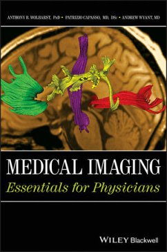
A well-known medical physicist and author, an interventional radiologist, and an emergency room physician with no special training in radiology have collaborated to write, in the language familiar to physicians, an introduction to the technology and clinical applications of medical imaging. It is intentionally brief and not overly detailed, intended to help clinicians with very little free time rapidly gain enough command of the critically important imaging tools of their trade to be able to discuss them confidently with medical and technical colleagues; to explain the general ideas accurately to students, nurses, and technologists; and to describe them effectively to concerned patients and loved ones. Chapter coverage includes:
- Introduction: Dr. Doe's Headaches
- Sketches of the Standard Imaging Modalities
- Image Quality and Dose
- Creating Subject Contrast in the Primary X-Ray Image
- Twentieth-Century (Analog) Radiography and Fluoroscopy
- Radiation Dose and Radiogenic Cancer Risk
- Twenty-First-Century (Digital) Imaging
- Digital Planar Imaging
- Computed Tomography
- Nuclear Medicine (Including SPECT and PET)
- Diagnostic Ultrasound (Including Doppler)
- MRI in One Dimension and with No Relaxation
- Mapping T1 and T2 Proton Spin Relaxation in 3D
- Evolving and Experimental Modalities
Dieser Download kann aus rechtlichen Gründen nur mit Rechnungsadresse in D ausgeliefert werden.
Produktdetails
- Produktdetails
- Verlag: Wiley
- Erscheinungstermin: 26. März 2013
- Englisch
- ISBN-13: 9781118480281
- Artikelnr.: 38247466
- Verlag: Wiley
- Erscheinungstermin: 26. März 2013
- Englisch
- ISBN-13: 9781118480281
- Artikelnr.: 38247466
- Herstellerkennzeichnung Die Herstellerinformationen sind derzeit nicht verfügbar.
Anthony Brinton Wolbarst, PhD, a physicist formerly at Harvard Medical School, the National Cancer Institute, and the U.S. Environmental Protection Agency, is currently an Associate Professor at the University of Kentucky College of Health Sciences, Division of Radiation Sciences and College of Medicine, Department of Diagnostic Radiology in Lexington, Kentucky, USA.
Patrizio Capasso, MD, is Professor and Division Chief of Vascular & Interventional Radiology in the Departments of Diagnostic Radiology and Surgery at the University of Kentucky Chandler Medical Center Lexington, Kentucky, USA.
Andrew R. Wyant, MD, is Assistant Professor for Physician Assistant Studies at the University of Kentucky Chandler Medical Center Lexington, Kentucky, USA. Among many other courses that he teaches is a popular clinical skills seminar in in Radiographic Interpretation.
Preface x
Acknowledgments xiii
Introduction: Dr. Doe's Headaches: An Imaging Case Study xiv
Computed tomography xiv
Picture archiving and communication system xv
T1, T2, and FLAIR MRI xvi
MR spectroscopy and a virtual biopsy xvii
Functional MRI xviii
Diffusion tensor MR imaging xviii
MR guided biopsy xx
Pathology xxi
Positron emission tomography? xxi
Treatment and follow-up xxii
1 Sketches of the Standard Imaging Modalities: Different Ways of Creating
Visible Contrast Among Tissues 1
"Roentgen has surely gone crazy!" 2
Different imaging probes interact with different tissues in different ways
and yield different kinds of medical information 4
Twentieth-century (analog) radiography and fluoroscopy: contrast from
differential attenuation of X-rays by tissues 7
Twenty-first century (digital) images and digital planar imaging:
computer-based images and solid-state image receptors 16
Computed tomography: three-dimensional mapping of X-ray attenuation by
tissues 17
Nuclear medicine, including SPECT and PET: contrast from the differential
uptake of a radiopharmaceutical by tissues 20
Diagnostic ultrasound: contrast from differences in tissue elasticity or
density 26
Magnetic resonance imaging: mapping the spatial distribution of
spin-relaxation times of hydrogen nuclei in tissue water and lipids 28
Appendix: selection of imaging modalities to assist in medical diagnosis 30
References 36
2 Image Quality and Dose: What Constitutes a "Good" Medical Image? 37
A brief history of magnetism 37
About those probes and their interactions with matter . . . 39
The image quality quartet: contrast, resolution, stochastic (random) noise,
artifacts - and always dose 47
Quality assurance 57
Known medical benefits versus potential radiation risks 61
3 Creating Subject Contrast in the Primary X-ray Image: Projection Maps of
the Body from Differential Attenuation of X-rays by Tissues 67
Creating a (nearly) uniform beam of penetrating X-rays 69
Interaction of X-ray and gamma-ray photons with tissues or an image
receptor 75
What a body does to the beam: subject contrast in the pattern of X-rays
emerging from the patient 83
What the beam does to a body: dose and risk 87
4 Twentieth-century (Analog) Radiography and Fluoroscopy: Capturing the
X-ray Shadow with a Film Cassette or an Image Intensifier Tube plus
Electronic Optical Camera Combination 91
Recording the X-ray pattern emerging from the patient with a screen-film
image receptor 92
Prime determinants/measures of image quality: contrast, resolution, random
noise, artifacts, . . . and, always, patient dose 98
Special requirements for mammography 114
Image intensifier-tube fluoroscopy: viewing in real time 122
Conclusion: bringing radiography and fluoroscopy into the twenty-first
century with solid-state digital X-ray image receptors 125
Reference 126
5 Radiation Dose and Radiogenic Risk: Ionization-Induced Damage to DNA can
cause Stochastic, Deterministic, and Teratogenic Health Effects - And How
To Protect Against Them 127
Our exposure to ionizing radiation has doubled over the past few decades
127
Radiation health effects are caused by damage to DNA 129
Stochastic health effects: cancer may arise from mutations in a single cell
132
Deterministic health effects at high doses: radiation killing of a large
number of tissue cells 139
The Four Quartets of radiation safety 146
References 151
6 Twenty-first Century (Digital) Imaging: Computer-Based Representation,
Acquisition, Processing, Storage, Transmission, and Analysis of Images 152
Digital computers 153
Digital acquisition and representation of an image 157
Digital image processing: enhancing tissue contrast, SNR, edge sharpness,
etc. 166
Computer networks: PACS, RIS, and the Internet 168
Image analysis and interpretation: computer-assisted detection 170
Computer and computer-network security 172
Liquid crystal displays and other digital displays 173
The joy of digital 174
7 Digital Planar Imaging: Replacing Film and Image Intensifiers with Solid
State, Electronic Image Receptors 176
Digital planar imaging modalities 176
Indirect detection with a fluorescent screen and a CCD 178
Computed radiography 178
Digital radiography with an active matrix flat panel imager 179
Digital mammography 184
Digital fluoroscopy and digital subtraction angiography 186
Digital tomosynthesis: planar imaging in three dimensions 189
References 190
8 Computed Tomography: Superior Contrast in Three-Dimensional X-Ray
Attenuation Maps 191
Computed tomography maps out X-ray attenuation in two and three dimensions
192
Image reconstruction 198
Seven generations of CT scanners 204
Technology and image quality 208
Patient- and machine-caused artifacts 219
Dose and QA 221
Appendix: mathematical basis of filtered back-projection 229
References 233
9 Nuclear Medicine: Contrast from Differential Uptake of a
Radiopharmaceutical by Tissues 234
Unstable atomic nuclei: radioactivity 235
Radiopharmaceuticals: gamma- or positron-emitting radionuclei attached to
organ-specific agents 245
Imaging radiopharmaceutical concentration with a gamma camera 248
Static and dynamic studies 254
Tomographic nuclear imaging: SPECT and PET 260
Quality assurance and radiation safety 270
References 273
10 Diagnostic Ultrasound: Contrast from Differences in Tissue Elasticity or
Density Across Boundaries 274
Medical ultrasound 274
The US beam: MHz compressional waves in tissues 277
Production of an ultrasound beam and detection of echoes with a transducer
280
Piezoelectric transducer elements 281
Transmission and attenuation of the beam within a homogeneous material 285
Reflection of the beam at an interface between materials with different
acoustic impedances 288
Imaging in 1 and 1 × 1 dimensions: A- and M-modes 291
Imaging in two, three, and four dimensions: B-mode 294
Doppler imaging of blood flow 300
Elastography 302
Safety and QA 303
11 MRI in One Dimension and with No Relaxation: A Gentle Introduction to a
Challenging Subject 307
Prologue to MRI 308
"Quantum" approach to proton nuclear magnetic resonance 310
Magnetic resonance imaging in one dimension 316
"Classical" approach to NMR 321
Free induction decay imaging (but without the decay) 331
Spin-echo imaging (still without T1 or T2 relaxation) 338
MRI instrumentation 343
Reference 351
12 Mapping T1 and T2 Relaxation in Three Dimensions 352
Longitudinal spin relaxation and T1 353
Transverse spin relaxation and T2-w images 364
T2¿ and the gradient-echo (G-E) pulse sequence 372
Into two and three dimensions 374
MR imaging of fluid movement/motion 382
13 Evolving and Experimental Modalities 387
Optical and near-infrared imaging 388
Molecular imaging and nanotechnology 390
Thermography 392
Terahertz (T-ray) imaging of epithelial tissues 393
Microwave and electron spin resonance imaging 393
Electroencephalography, magnetocardiography, and impedance imaging 394
Photo-acoustic imaging 396
Computer technology: the constant revolution 397
Imaging with a crystal ball 399
References 399
Suggested Further Reading 400
Index 403
