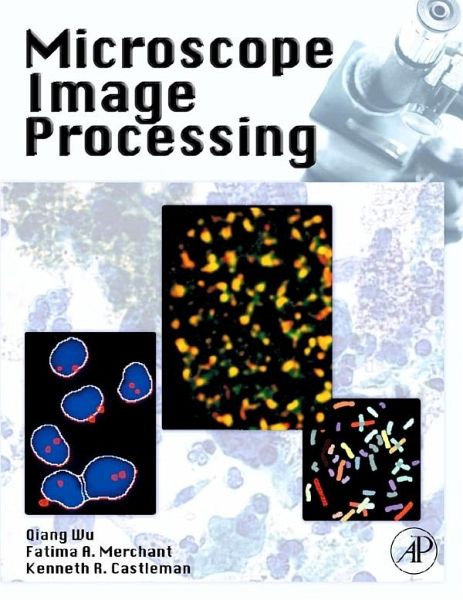
Digital image processing, an integral part of microscopy, is increasingly important to the fields of medicine and scientific research. This book provides a unique one-stop reference on the theory, technique, and applications of this technology.
Written by leading experts in the field, this book presents a unique practical perspective of state-of-the-art microscope image processing and the development of specialized algorithms. It contains in-depth analysis of methods coupled with the results of specific real-world experiments. Microscope Image Processing covers image digitization and display, object measurement and classification, autofocusing, and structured illumination.
Key Features:
This book is of interest to all scientists, engineers, clinicians, post-graduate fellows, and graduate students working in the fields of biology, medicine, chemistry, pharmacology, and other related fields. Anyone who uses microscopes in their work and needs to understand the methodologies and capabilities of the latest digital image processing techniques will find this book invaluable.
Written by leading experts in the field, this book presents a unique practical perspective of state-of-the-art microscope image processing and the development of specialized algorithms. It contains in-depth analysis of methods coupled with the results of specific real-world experiments. Microscope Image Processing covers image digitization and display, object measurement and classification, autofocusing, and structured illumination.
Key Features:
- Detailed descriptions of many leading-edge methods and algorithms
- In-depth analysis of the method and experimental results, taken from real-life examples
- Emphasis on computational and algorithmic aspects of microscope image processing
- Advanced material on geometric, morphological, and wavelet image processing, fluorescence, three-dimensional and time-lapse microscopy, microscope image enhancement, MultiSpectral imaging, and image data management
This book is of interest to all scientists, engineers, clinicians, post-graduate fellows, and graduate students working in the fields of biology, medicine, chemistry, pharmacology, and other related fields. Anyone who uses microscopes in their work and needs to understand the methodologies and capabilities of the latest digital image processing techniques will find this book invaluable.
- Presents a unique practical perspective of state-of-the-art microcope image processing and the development of specialized algorithms
- Each chapter includes in-depth analysis of methods coupled with the results of specific real-world experiments
- Co-edited by Kenneth R. Castleman, world-renowned pioneer in digital image processing and author of two seminal textbooks on the subject
Dieser Download kann aus rechtlichen Gründen nur mit Rechnungsadresse in A, B, BG, CY, CZ, D, DK, EW, E, FIN, F, GR, HR, H, IRL, I, LT, L, LR, M, NL, PL, P, R, S, SLO, SK ausgeliefert werden.
"Such a book is much-needed!!!" --Al Bovik, ECE Dept. Head, Univ. of Texas; Editor of Handbook of Image and Video Processing (AP 2005)
"My impression is that this book would be very useful and approachable to microscopists/biologists and to image processing specialists working with them. ... I would suspect a high market potential." --Jeff Pierce, PhD, R&D Staff Member, Image Science and Machine Vision Group, Oak Ridge National Lab
"The authors propose to write a book...[with] a good balance between theoretical framework and practical applications.... The authors are certainly qualified for the task. In particular, Dr. Castleman's expertise and recognition in this arena will likely evoke good contributions from leading experts in the field." --Dr. Alberto Goldszal, U. of PA Health System, Dir. of Medical Informatics, Dept. of Radiology
"I believe [there is] a large community who will likely find a book like this to be of technical relevance to their work and a good reference.... The concept of focusing on the image processing applications in microscopy is good and i do think that this will make the resource valuable." --Kenneth Tobin, Oak Ridge Nat'l Lab, Group Leader/Corporate Research Fellow
"It would be very helpful to have a practical guide to digital image processing since much of the material is currently embedded in software program guides available only with the purchase of high end graphics programs or in technical journals not generally read by biologists, biochemists, and other emerging users of advanced optical imaging techniques. The technical soundness of the proposal is well thought-out." --Dr. Paul Kulesa, Dir. of Imaging, Stowers Inst. for Medical Research