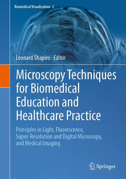
Microscopy Techniques for Biomedical Education and Healthcare Practice (eBook, PDF)
Principles in Light, Fluorescence, Super-Resolution and Digital Microscopy, and Medical Imaging
Redaktion: Shapiro, Leonard
Versandkostenfrei!
Sofort per Download lieferbar
120,95 €
inkl. MwSt.
Weitere Ausgaben:

PAYBACK Punkte
60 °P sammeln!
This edited book has a strong focus on advances in microscopy that straddles research, medical education and clinical practice. These advances include the shift in power from conventional to digital microscopy. The first section of this book covers imaging techniques and morphometric image analysis with its applications in biomedicine using different microscopy modes. Chapters highlight the rich development of fluorescence methods and technologies; particle tracking techniques with applications in biomedical research and nanomedicine; the way in which visualizations have revolutionized taxonom...
This edited book has a strong focus on advances in microscopy that straddles research, medical education and clinical practice. These advances include the shift in power from conventional to digital microscopy. The first section of this book covers imaging techniques and morphometric image analysis with its applications in biomedicine using different microscopy modes. Chapters highlight the rich development of fluorescence methods and technologies; particle tracking techniques with applications in biomedical research and nanomedicine; the way in which visualizations have revolutionized taxonomy from gross anatomy to genetics; and the psychology of perception and how it affects our understanding of cells and tissues. The book's first section concludes by exploring the use of CT modalities to evaluate anterior deformities in craniosynostosis. In the second section of the book, chapters on anatomical and cell biology education explore the history of anatomical models and their use in educational settings. This includes examples in 3D printing and functional human anatomical models that can be created using easily available resources and the use of biomedical imaging in visuospatial teaching of anatomy; the novel use of ultrasound in medical education and practice; and skill acquisition in histology education using a flowchart called a 'decision tree'.
This book will appeal to histologists, microscopists, cell biologists, clinicians and those involved in anatomical education and biomedical visualization, as well as students in those respective fields.
This book will appeal to histologists, microscopists, cell biologists, clinicians and those involved in anatomical education and biomedical visualization, as well as students in those respective fields.
Dieser Download kann aus rechtlichen Gründen nur mit Rechnungsadresse in A, B, BG, CY, CZ, D, DK, EW, E, FIN, F, GR, HR, H, IRL, I, LT, L, LR, M, NL, PL, P, R, S, SLO, SK ausgeliefert werden.


